
NORCET-11 Mains
Medical & Surgical Nursing
Medium
A 42-year-old client presents with acute chest pain. Which type of myocardial infarction is shown on the ECG? 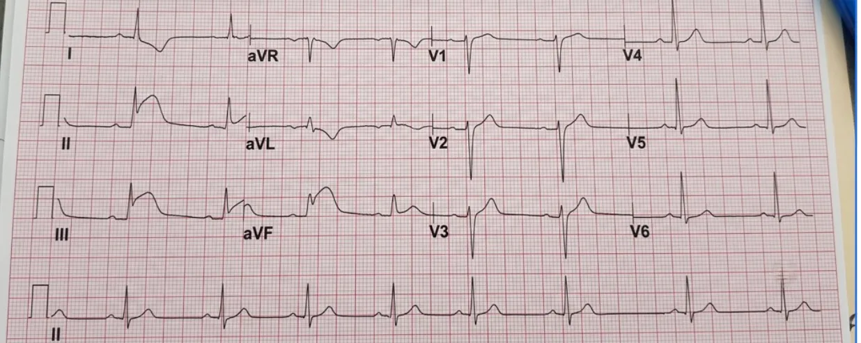
Appeared in: NORCET-11 Mains
Explanation
- The ECG shows ST-segment elevation in leads II, III, and aVF.
- These leads (II, III, aVF) directly view the inferior wall of the left ventricle.
- ST-segment elevation in this specific grouping is the hallmark of an acute injury to the inferior myocardial wall.
- The presence of reciprocal ST depression in leads I and aVL confirms the diagnosis of an inferior MI.
Why Other Options Were Wrong
- Option A: An anterior wall MI would present with ST-segment elevation in the precordial leads, specifically V1 through V4. The provided ECG does not show these changes.
- Option B: A lateral wall MI would show ST-segment elevation in the lateral leads (I, aVL, V5, V6). In this ECG, leads I and aVL actually show ST depression, which are reciprocal changes to the inferior injury.
- Option D: A posterior wall MI is typically identified by indirect (reciprocal) changes in the anterior leads (V1-V3), such as ST depression and tall R waves. While some ST depression might be present, the primary and most obvious finding is the ST elevation in the inferior leads.
Related Visual

Clinical Relevance
- Nursing practice connection: This is primarily an exam-oriented knowledge point with limited direct bedside application, so retain ECG Interpretation for Myocardial Infarction Localization as background academic context rather than a clinical decision trigger.
- Rapid identification of the MI location is critical for predicting potential complications. Inferior MIs are often associated with bradycardia and heart blocks because the Right Coronary Artery (RCA), which is usually the culprit, also supplies the SA and AV nodes.
- Nurses must be prepared to manage hypotension and bradycardia in patients with inferior MI. Atropine and temporary pacing may be required.
- It is crucial to obtain a right-sided ECG (e.g., lead V4R) in patients with inferior MI to assess for right ventricular involvement, as this significantly impacts treatment (e.g., nitrates should be used with extreme caution or avoided).
How to Approach the Question
- First, identify that the question requires interpretation of a 12-lead ECG to locate a myocardial infarction.
- Systematically scan all 12 leads, looking for the most significant abnormality. The key finding in an acute MI is ST-segment elevation.
- Observe that there is clear ST-segment elevation in leads II, III, and aVF.
- Recall or reference the standard ECG lead groupings. Leads II, III, and aVF correspond to the inferior wall of the heart.
- Look for reciprocal changes to confirm the finding. ST depression in leads I and aVL is the expected reciprocal pattern for an inferior MI.
- Based on the ST elevation in the inferior leads, conclude that the patient is having an inferior wall myocardial infarction and select the corresponding option.
Concept Tested & Keywords
- Concept Tested: ECG Interpretation for Myocardial Infarction Localization
- Stem keywords: ECG, acute chest pain, myocardial infarction
- Lead-in keywords: Which type
- Clinical cues: The presence of ST-segment elevation in leads II, III, and aVF is the key diagnostic finding.
Question ID
QBhNscmXR00ONQ-_lfIX7i
Reference Book
E6 Medicine Harrison 22e Part 1 pp. 1913-1915, 1914-1916
Practise the full NORCET-11 Mains
Attempt every question from this paper in a timed mock, then review the full solution for each one.
More Cardiovascular, Circulatory, and Hematologic Function Questions
- A patient was brought to ED with complaint of chest pain from last 1 hour. Which test is done to detect myocardial infarction?
- A patient is undergoing blood transfusion and suddenly shows complaints of transfusion reaction. What will be the priority nursing intervention?
- Normal hemoglobin level in adult is:
- The following all are blood transfusion complications from; EXCEPT:
- An obstruction of blood flowing from the left atrium into the left ventricle is known as:
- 'Sequestration crisis' is related to:
- Calculate the pulse pressure from the below image of cardiac monitor:
- Identify the use of following machine used in pneumatic compression device:
- Identify the red wave forms in the following image:
- Identify the catheter in the following image:
More NORCET-11 Mains Questions
- A postoperative day 1 client after coronary artery bypass grafting has a sudden decrease in drain output from 50 mL to 0 mL, hypotension, and distend…
- A client with atrial fibrillation suddenly develops a heart rate of 175 to 195 per minute, blood pressure of 80/50 mmHg, chest pain, dizziness, and o…
- A patient underwent femoral artery catheterization. After the procedure, the affected leg becomes numb and pale, with decreased circulation and a wea…
- A client is brought unconscious with pinpoint pupils and a respiratory rate of 8 per minute. What is the first priority?
- A known diabetic client comes to the emergency department with a random blood sugar of 400 mg/dL, Kussmaul respirations, and a serum potassium of 5.8…
- A client passes 3.5 liters of urine a day. Plasma osmolality is 305 mOsm/kg, urine osmolality is 150 mOsm/kg, urine specific gravity is 1.000, and pl…
- A client in the ward suddenly develops epistaxis and feels blood moving toward the throat. To prevent aspiration, which position should the client be…
- A client comes in with suspected acute cholecystitis. Which abdominal finding most strongly supports the diagnosis?
- Which test is performed for a suspected meniscal tear by flexing the knee and rotating the tibia while extending the knee?
- Identify the traction shown, used for a femur fracture in a child under 2 years.& x20;