
NORCET Mains-11
Medical & Surgical Nursing
Hard
A road traffic accident victim has absent breath sounds and tracheal deviation. The X-ray shows a hyperlucent hemithorax without lung markings and mediastinal shift to the opposite side. What is the diagnosis?
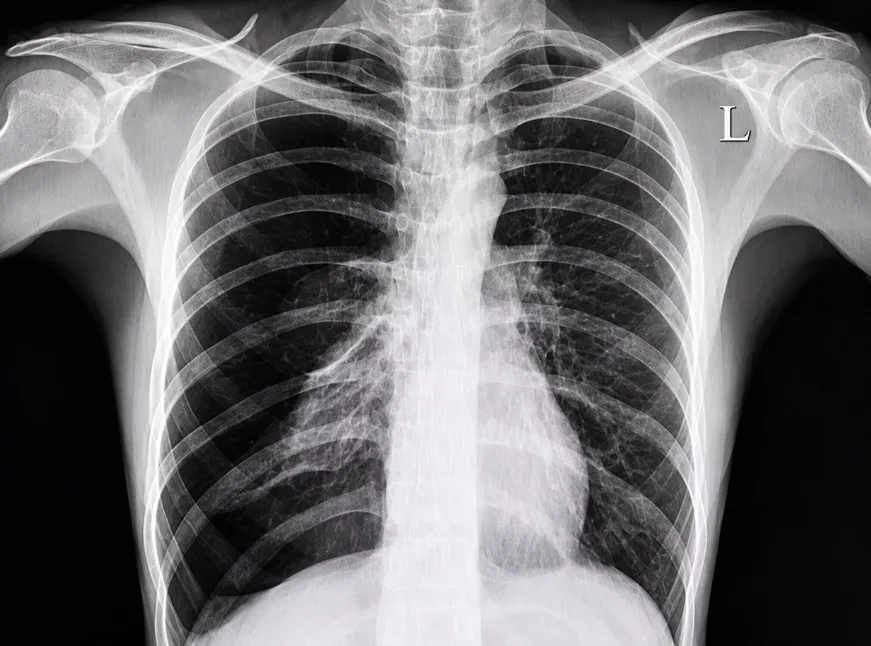
Appeared in: NORCET Mains-11
Explanation
- Tension pneumothorax is a life-threatening condition where a one-way valve effect allows air to enter the pleural space but not escape.
- The accumulating air increases pressure, which collapses the lung on the affected side (causing absent breath sounds) and pushes the trachea and heart to the opposite side (causing contralateral mediastinal shift).
- The chest X-ray confirms the diagnosis by showing a hyperlucent (black) hemithorax due to the trapped air, absence of lung markings from the collapsed lung, and a clear shift of the mediastinum away from the affected side.
Why Other Options Were Wrong
- Option B: Massive hemothorax is the accumulation of blood, not air, in the pleural space. On an X-ray, this would appear as opacification (whiteness or a 'white-out'), which is the opposite of the hyperlucency (blackness) described.
- Option C: Flail chest is a structural injury of the chest wall involving multiple rib fractures, leading to paradoxical chest movement. While it can cause a pneumothorax, the defining feature is the unstable chest wall segment, not the specific combination of hyperlucency and mediastinal shift that defines tension.
- Option D: Pleural effusion is a collection of fluid (e.g., serous fluid, pus) in the pleural space. Like hemothorax, it appears as an opacification (white) on an X-ray, often with a characteristic curved upper border (meniscus sign) and blunting of the costophrenic angle.
Related Visual

Clinical Relevance
- Nursing practice connection: Use the key finding related to Diagnosis of tension pneumothorax using clinical and radiological findings to guide bedside assessment, documentation, and the next nursing action.
- Tension pneumothorax is a critical, life-threatening emergency. Nurses must be able to recognize the signs (respiratory distress, unilateral absent breath sounds, tracheal deviation, hypotension) for immediate action.
- The priority intervention is to alert the physician and prepare for an emergency needle decompression (thoracentesis) in the second intercostal space, midclavicular line, followed by chest tube insertion.
- Failure to decompress a tension pneumothorax can lead to obstructive shock and cardiovascular collapse within minutes.
How to Approach the Question
- First, analyze the clinical context: a road traffic accident victim implies a high likelihood of thoracic trauma.
- Next, identify the key clinical signs presented: absent breath sounds on one side combined with tracheal deviation to the opposite side. This pairing is a classic indicator of a pressure-building lesion in the chest.
- Then, interpret the radiological findings. 'Hyperlucent' means excess air. 'Absent lung markings' means the lung is collapsed. 'Mediastinal shift to the opposite side' confirms that pressure is pushing structures away.
- Evaluate each option against this complete clinical and radiological picture. Tension pneumothorax is the only diagnosis that accounts for all these specific findings.
- Eliminate other options based on their expected X-ray appearance: hemothorax and pleural effusion would cause opacification (whiteness), not hyperlucency.
Concept Tested & Keywords
- Concept Tested: Diagnosis of tension pneumothorax using clinical and radiological findings.
- Stem keywords: road traffic accident, absent breath sounds, tracheal deviation, hyperlucent hemithorax, absent lung markings, mediastinal shift
- Lead-in keywords: What is the diagnosis?
- Clinical cues: Tracheal deviation away from the side with absent breath sounds is a critical sign of tension pneumothorax.
- Clinical cues: Hyperlucency on a chest X-ray indicates the presence of air in a space where it shouldn't be, such as the pleural cavity.
Question ID
QIIUQaK_kTxUtTFHJ8DKrR
Reference Book
E6 Nursing Brunner Adult Health 3SA Vol 1 Part 2 p. 24-26
E6 Nelson Textbook of Pediatrics(2024) — Volume 2 p. 492-494
Practise the full NORCET Mains-11
Attempt every question from this paper in a timed mock, then review the full solution for each one.
More Gas Exchange and Respiratory Function Questions
- Which is the most relevant knowledge about oxygen administration to a client with COPD?
- Patient in emergency department diagnosed to have fluid and air present in lung. What is the above-mentioned condition known as?
- Patient is suffering from dyspnea. Which position will you provide for patient to relieve?
- Which of the following is not involved in chest physiotherapy?
- Respiration which is abnormally shallow for two or three breaths followed by irregular period of apnea is termed:
- Identify the following instrument which is used in diagnostic procedures:
- What is the procedure shown in the image?
- Identify the instrument shown in the following image:
- Identify the tube in the following image:
- The device used to maintain the patency of the oral airways:
More NORCET Mains-11 Questions
- A 42-year-old client presents with acute chest pain. Which type of myocardial infarction is shown on the ECG?& x20;
- A postoperative day 1 client after coronary artery bypass grafting has a sudden decrease in drain output from 50 mL to 0 mL, hypotension, and distend…
- A client with atrial fibrillation suddenly develops a heart rate of 175 to 195 per minute, blood pressure of 80/50 mmHg, chest pain, dizziness, and o…
- A patient underwent femoral artery catheterization. After the procedure, the affected leg becomes numb and pale, with decreased circulation and a wea…
- A client is on postoperative day 5 after coronary artery bypass grafting. Vital signs are temperature 39 degrees Celsius, heart rate 130 per minute i…
- A patient on a mechanical ventilator has a low-pressure alarm. Oxygen saturation is falling and respiratory rate support is not being maintained. Ini…
- A polytrauma client on mechanical ventilation develops a high-pressure alarm, airway secretions, and oxygen saturation falls to 82 percent. What is t…
- A client is brought unconscious with pinpoint pupils and a respiratory rate of 8 per minute. What is the first priority?
- A known diabetic client comes to the emergency department with a random blood sugar of 400 mg/dL, Kussmaul respirations, and a serum potassium of 5.8…
- A client with diabetic ketoacidosis has a serum potassium of 3.0 mEq/L. The physician prescribes an insulin infusion and IV potassium. What should th…