
AIIMS Delhi NO- 2019
Medical & Surgical Nursing
Easy
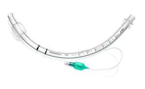
Identify the tube in the following image:
Appeared in: AIIMS Delhi NO- 2019
Explanation
- The image shows a flexible tube with an inflatable cuff at the tip, a pilot balloon for inflation, and depth markings, which are all characteristic features of an endotracheal tube (ETT).
- The primary purpose of an ETT is to secure a definitive airway for mechanical ventilation, administer oxygen, and protect the lungs from aspiration.
- The 15mm universal connector at the proximal end allows it to be connected to various respiratory devices like a ventilator or an Ambu bag.
- The inflatable cuff, when filled with air via the pilot balloon, creates a seal against the tracheal wall, preventing air leaks and aspiration of foreign materials.
Why Other Options Were Wrong
- Option A: An oropharyngeal airway (OPA) is a rigid, J-shaped device without a cuff or pilot balloon. It is placed in the mouth to keep the tongue from obstructing the airway, not inserted into the trachea.
- Option C: A tracheostomy tube is shorter and more curved than an ETT. It is inserted through a surgical opening (stoma) in the neck and has a flange that sits on the skin. The device in the image is long and designed for oral or nasal insertion.
- Option D: A Combi tube is a more complex, dual-lumen emergency airway device with two separate cuffs and two inflation ports. The tube in the image is a single-lumen device.
Related Visual

Clinical Relevance
- Nursing practice connection: This is primarily an exam-oriented knowledge point with limited direct bedside application, so retain Identification of Airway Management Devices as background academic context rather than a clinical decision trigger.
- Nurses are responsible for monitoring patients with endotracheal tubes, which includes assessing for correct placement, monitoring cuff pressure to prevent tracheal injury (necrosis or fistula), providing regular oral care to prevent ventilator-associated pneumonia (VAP), and ensuring the tube is secure.
- Communication is a key challenge, as intubated patients cannot speak. Nurses must establish alternative communication methods like whiteboards, picture boards, or simple yes/no questions.
- Emergency equipment, including a spare ETT, a laryngoscope, and a manual resuscitation bag (Ambu bag), must be kept at the bedside in case of accidental extubation or tube malfunction.
How to Approach the Question
- First, carefully examine the image provided, paying close attention to the specific features of the medical device.
- Identify key components: Is it a simple or complex tube? Does it have a cuff? Is there a pilot balloon? What is its shape and length? Note the presence of any connectors or markings.
- Systematically compare these observed features with the descriptions of the options provided.
- Eliminate options that do not match your visual analysis. For example, an oropharyngeal airway is uncuffed and rigid, which is different from the image.
- A tracheostomy tube has a neck flange, which is absent here. A Combi tube has two lumens, unlike the single lumen shown.
- Select the option that perfectly matches all the identified features. The cuffed, single-lumen tube with a pilot balloon is definitively an endotracheal tube.
Concept Tested & Keywords
- Concept Tested: Identification of Airway Management Devices
- Stem keywords: Identify the tube, image
- Lead-in keywords: BEST, MOST RELEVANT CLUE
Question ID
Q9fo4HXcoSVgsv8DV49HhY
Reference Book
E6 Nursing Fundamentals Taylor pp. 734-736, 736-738
Practise the full AIIMS Delhi NO- 2019
Attempt every question from this paper in a timed mock, then review the full solution for each one.
More Gas Exchange and Respiratory Function Questions
- Which is the most relevant knowledge about oxygen administration to a client with COPD?
- Patient in emergency department diagnosed to have fluid and air present in lung. What is the above-mentioned condition known as?
- Patient is suffering from dyspnea. Which position will you provide for patient to relieve?
- Which of the following is not involved in chest physiotherapy?
- Respiration which is abnormally shallow for two or three breaths followed by irregular period of apnea is termed:
- Identify the following instrument which is used in diagnostic procedures:
- Identify the instrument shown in the following image:
- A patient suddenly develops respiratory difficulties such as dyspnea, what is the first priority of the nursing officer?
- Which of the following method is used for high flow oxygen administration:
- Which of the following is the cardinal sign of pulmonary edema?
More AIIMS Delhi NO- 2019 Questions
- A nurse is preparing to give bath to admitted patient with perineal problem. Which of the following will help the patient?
- In the absence of nurse from floor, patient falls from bed. This type of injury belongs to:
- Patient's Medical record with all essential information is responsibility of:
- Post-partum women having bleeding within 24 hrs after delivery is called:
- In which condition, distal pulse is preferred rather than apical pulse?
- A pre-eclampsia woman's baby is lying with her mother. Neonate is born in 37 weeks. After 57 hours, when nurse is monitoring vital signs of baby, tem…
- A woman with 3rd day postpartum has complaint of engorgement in both breasts. What is cut off temperature for fever in postpartum women?
- Universal blood donor?
- WHO hand wash time with soap?
- Why it is necessary to remove dark coloured nail paint before surgery?