
NORCET 5 Prelims - Sept 2023 (Shift-1)
Medical & Surgical Nursing
Medium
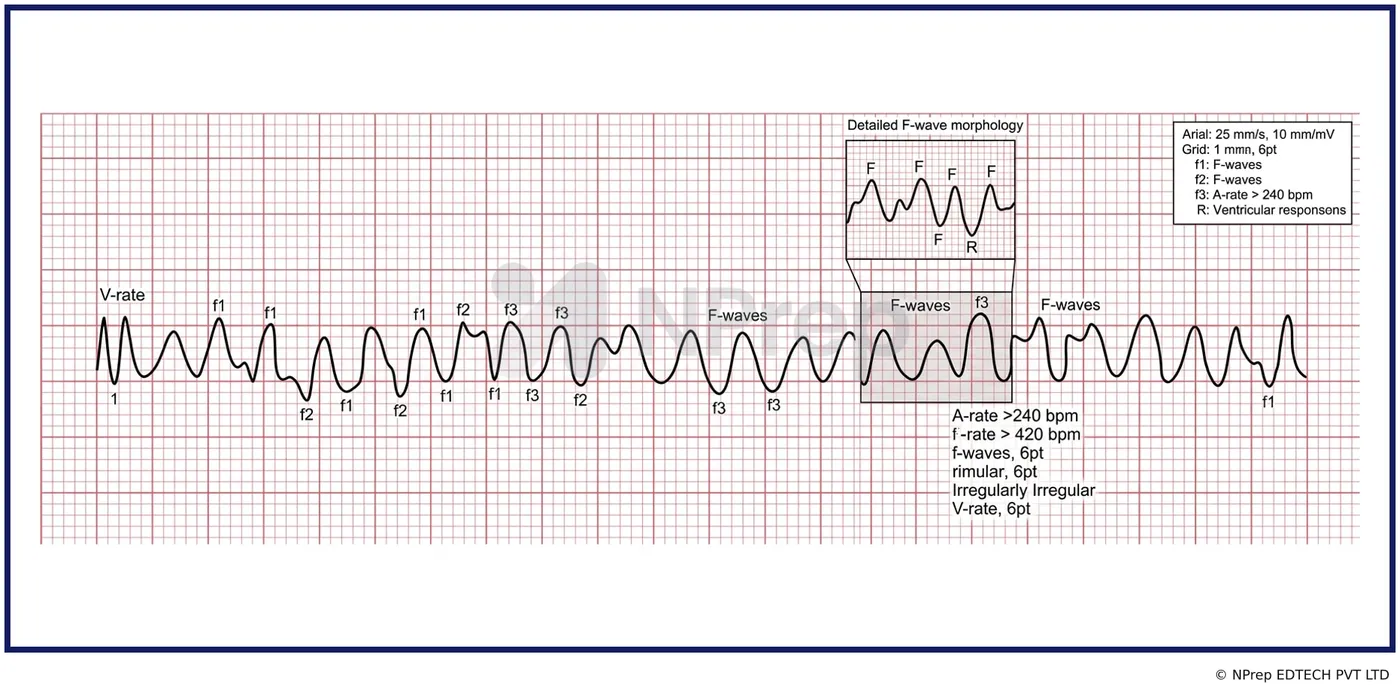
Identify the following ECG finding in the depicted image?
Appeared in: NORCET 5 Prelims - Sept 2023 (Shift-1)
Explanation
- The ECG demonstrates a completely chaotic and irregular waveform, which is the defining characteristic of Ventricular Fibrillation (VF).
- In this rhythm, there are no identifiable P waves, QRS complexes, or T waves; the baseline is replaced by fibrillatory waves of varying sizes and shapes.
- VF represents disorganized electrical activity in the ventricles, causing them to quiver instead of contracting, resulting in no cardiac output.
- This is a pulseless, life-threatening arrhythmia that requires immediate defibrillation and CPR.
Why Other Options Were Wrong
- Option B: Ventricular Tachycardia (VT) is typically a more organized rhythm, characterized by a rapid, regular (or sometimes slightly irregular) rhythm with wide QRS complexes. The ECG in the image is completely chaotic, not organized.
- Option C: Supraventricular Tachycardia (SVT) originates above the ventricles and is characterized by a rapid, regular rhythm with narrow QRS complexes. The ECG shown has no discernible QRS complexes and is chaotic, which is inconsistent with SVT.
- Option D: Atrial Fibrillation (AF) is characterized by an 'irregularly irregular' rhythm with no P waves but with identifiable (usually narrow) QRS complexes. The ECG in the image lacks any identifiable QRS complexes and shows complete ventricular chaos, which is far more severe than AF.
Related Visual

Clinical Relevance
- Nursing practice connection: This is primarily an exam-oriented knowledge point with limited direct bedside application, so retain ECG interpretation of lethal cardiac arrhythmias as background academic context rather than a clinical decision trigger.
- Rapid recognition of Ventricular Fibrillation on a cardiac monitor is a critical nursing skill. It is the most common initial rhythm in out-of-hospital cardiac arrest.
- Immediate initiation of the 'Code Blue' or emergency response protocol, including high-quality CPR and defibrillation, is the only effective treatment.
- A nurse's primary role is to confirm pulselessness, call for help, start CPR, and prepare for defibrillation as per ACLS guidelines.
How to Approach the Question
- First, assess the overall appearance of the ECG strip. Note if it is organized or chaotic.
- Second, attempt to identify key components: Is there a regular rhythm? Are there P waves? Are the QRS complexes present, and are they narrow or wide?
- In this image, observe that the entire waveform is disorganized and chaotic, with no identifiable components. This immediately points towards a fibrillatory rhythm.
- Next, determine the origin. Since the chaos involves the entire complex and represents a non-perfusing state, it originates in the ventricles.
- Based on the chaotic, irregular, and non-perfusing nature with no discernible QRS complexes, conclude the rhythm is Ventricular Fibrillation.
- Eliminate other options: VT is more organized, while SVT and AF have identifiable QRS complexes.
Concept Tested & Keywords
- Concept Tested: ECG interpretation of lethal cardiac arrhythmias.
- Stem keywords: ECG finding, depicted image
- Lead-in keywords: Identify
- Clinical cues: The ECG shows a chaotic, disorganized rhythm, which is a critical visual cue.
Question ID
QFvn1G0lk6ayn9kGRsEzrM
Reference Book
E6 Medicine Harrison 22e Part 1 p. 1996-1998
E6 Nelson Textbook of Pediatrics(2024) — Volume 2 p. 642-644
Practise the full NORCET 5 Prelims - Sept 2023 (Shift-1)
Attempt every question from this paper in a timed mock, then review the full solution for each one.
More Cardiovascular, Circulatory, and Hematologic Function Questions
- A patient is undergoing blood transfusion and suddenly shows complaints of transfusion reaction. What will be the priority nursing intervention?
- The following all are blood transfusion complications from; EXCEPT:
- Identify the use of following machine used in pneumatic compression device:
- In iron deficiency anemia, the fingernails become spoon shaped, this condition is referred to as:
- Which gauze cannula is commonly used for blood transfusion?
- If 'P' wave is absent in ECG rhythm, it is an indication of:
- Normal compression to ventilation ratio:
- Trans-illumination procedure is done with:
- A patient in the ward experiences sudden cardiac arrest. What is the most appropriate technique for opening the airway to facilitate endotracheal int…
- A patient experiences complications after a blood transfusion. Which of the following signs and symptoms is indicative of circulatory overload?
More NORCET 5 Prelims - Sept 2023 (Shift-1) Questions
- Which of the following electrocardiogram finding would be seen in a client with Hyperkalemia?
- Which of the following is not used in management of orthostatic hypotension?
- Which of the following clinical features is not seen in shock?
- Which position is best to identify left pleural effusion?
- A male patient comes in casualty who has a lacerated untidy wound after 4 hours. What would be the priority nursing action?
- A client who underwent abdominal surgery earlier in the day has a small amount of bright red blood on the surgical dressing. How should the nurse doc…
- A nurse entered a nursery unit and observed that a newborn shows jerky movement of the lower limbs. Which of the following is the priority nursing ac…
- The nurse should be concerned for which while a child is on chemotherapy?
- Following a head injury, a person comes into the emergency department with personality changes and loss of voluntary control. Which lobe of the brain…
- Which of the following solution is only used for dilution of phenytoin (Dilantin)?