
Combined AIIMS - 2020 (Jodhpur, Bhopal, Patna, Raipur)
Nursing Research & Statistics
Medium
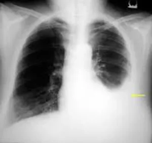
Identify the disorder presented in X-ray:
Appeared in: Combined AIIMS - 2020 (Jodhpur, Bhopal, Patna, Raipur)
Explanation
- The X-ray shows a dense, uniform opacity in the left lower lung field, which is characteristic of fluid accumulation.
- A key finding is the blunting or obscuring of the left costophrenic angle, a classic sign of pleural effusion.
- The concave upper border of the opacity, known as the meniscus sign, confirms the presence of fluid in the pleural space.
- These combined features are the hallmark radiological presentation of a pleural effusion.
Why Other Options Were Wrong
- Option B: Right-sided pneumonia would present as a consolidation or infiltrate within the lung tissue on the right side, often with air bronchograms. The image shows a fluid collection outside the lung on the left side.
- Option C: Pericarditis itself is not visible on a standard X-ray. If it leads to a significant pericardial effusion, the heart silhouette may appear enlarged and globular (like a 'water bottle'), which is not the case here. The abnormality is in the pleural space, not the pericardial sac.
- Option D: Bronchiectasis involves the permanent dilation of airways. On an X-ray, this appears as thickened bronchial walls, seen as parallel 'tram tracks' or ring shadows. These findings are not present in the given image.
Related Visual

Clinical Relevance
- Nursing practice connection: This is primarily an exam-oriented knowledge point with limited direct bedside application, so retain Radiological interpretation of common thoracic disorders, specifically pleural effusion as background academic context rather than a clinical decision trigger.
- Recognizing pleural effusion on an X-ray is a critical skill for nurses to anticipate patient needs and potential interventions.
- A nurse observing these signs should prioritize respiratory assessment, including oxygen saturation, work of breathing, and lung sounds (which would be diminished or absent over the effusion).
- The nurse should prepare for potential diagnostic or therapeutic procedures, such as a thoracentesis (pleural tap), by ensuring informed consent is obtained and necessary equipment is available.
How to Approach the Question
- First, systematically examine the chest X-ray. Check the airway, bones, cardiac silhouette, diaphragm, and lung fields (the 'ABCDE' approach).
- Focus on the lung bases and costophrenic angles. Note that the right angle is sharp, while the left is obscured.
- Identify the key abnormal finding: a dense opacity at the left lung base with a concave upper border.
- Correlate this finding with the classic radiological signs of the conditions listed in the options.
- Pleural effusion is defined by fluid blunting the costophrenic angle and forming a meniscus, which directly matches the image.
- Rule out the other options by confirming their characteristic signs (e.g., consolidation for pneumonia, tram tracks for bronchiectasis) are absent.
Concept Tested & Keywords
- Concept Tested: Radiological interpretation of common thoracic disorders, specifically pleural effusion.
- Stem keywords: X-ray, disorder, chest
- Lead-in keywords: Identify
- Clinical cues: The image is a chest X-ray showing abnormal findings in the left lung base.
Question ID
QKO5KbZ3ZEub_uqLfaq5Xe
Reference Book
E6 Medicine Davidson Principles Practice 24e p. 583-585
Practise the full Combined AIIMS - 2020 (Jodhpur, Bhopal, Patna, Raipur)
Attempt every question from this paper in a timed mock, then review the full solution for each one.
More Communication and utilization of Research Questions
- Which mode of clinical application for qualitative research is considered to be the sharing of qualitative findings with the patient?
- What is the term used for a careful appraisal of the strengths and weakness of a research study?
- A clear, concise summary of a study that communicates the essential information about the study is called?
- Which guideline is used for writing a report of a randomized control trial research study?
- Comparing the present study finding with existing literature is included in?
- The process of application of the research findings done on the nurses in Hyderabad to all nurses in Telangana is known as?
- The following is an example of a visual graphic presentation of study results?
- A nurse who reads research articles and incorporates research findings into nursing practice would demonstrate one of the following research roles :
- The researcher uses one of the following portions of the research report to capture the reader's attention :
- In the context of utilizing research in nursing practice, what is the primary role of clinical guidelines derived from systematic reviews?& x20;
More Combined AIIMS - 2020 (Jodhpur, Bhopal, Patna, Raipur) Questions
- The position used for most of the gynecological procedures?
- Thyroid eye disease (TED) is also known as:
- How many items are included in the WHO surgical safety checklist: SITOSO?
- In iron deficiency anemia, the fingernails become spoon shaped, this condition is referred to as:
- Which of the following code is associated with cardiac arrest during medical emergency?
- How is blood loss measured in Post-partum hemorrhage?
- The traction used for cervical spine injury:
- In operation theater during cauterization, the electric current passing from a single electrode is called:
- Tracheostomy is done in which tracheal ring?
- The instrument used for measuring fetal heart rate (FHR):