
NORCET 7 Prelims -2024
Medical & Surgical Nursing
Easy
Based on the image showing the placement of ECG electrodes, where should the V1 lead be placed?
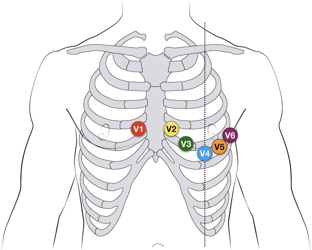
Appeared in: NORCET 7 Prelims -2024
Explanation
- The V1 lead is the first of the six precordial (chest) leads in a standard 12-lead ECG.
- Its correct anatomical position is in the fourth intercostal space (the space below the 4th rib).
- This position is located immediately to the right of the sternum (breastbone).
- This placement allows the V1 lead to record electrical activity from the right ventricle and the interventricular septum.
Why Other Options Were Wrong
- Option B: This describes the correct placement for the V4 lead, not the V1 lead.
- Option C: This describes the correct placement for the V6 lead, not the V1 lead.
- Option D: This describes the correct placement for the V2 lead. It is a common point of confusion as it is in the same intercostal space as V1 but on the opposite side of the sternum.
Related Visual

Clinical Relevance
- Nursing practice connection: This is primarily an exam-oriented knowledge point with limited direct bedside application, so retain Standard placement of precordial (chest) leads for a 12-lead electrocardiogram (ECG) as background academic context rather than a clinical decision trigger.
- Accurate ECG lead placement is a fundamental nursing skill and is non-negotiable for diagnostic accuracy. Misplacing leads can create artifactual ECG changes that mimic serious conditions like myocardial infarction, bundle branch block, or ventricular hypertrophy, leading to incorrect diagnosis and inappropriate treatment.
- For example, placing V1 and V2 leads too high (in the 2nd or 3rd intercostal space) is a common error that can produce a pattern resembling an anterior myocardial infarction (Brugada pattern), potentially causing unnecessary anxiety and further invasive testing.
- What if V1 and V2 are accidentally swapped? Swapping V1 and V2 results in a loss of the normal R-wave progression across the precordium. The R-wave in the new 'V1' (actually V2) will be larger than expected, and the R-wave in the new 'V2' (actually V1) will be smaller, which can mask or mimic septal and anterior wall abnormalities.
How to Approach the Question
- First, identify the core question being asked, which is the specific anatomical location for the 'V1 lead'.
- Use the provided image as a visual reference. The image confirms the question is about the standard precordial (chest) lead setup.
- Systematically recall the anatomical landmarks for the six V-leads. A good strategy is to remember them in order.
- Start with V1: Locate the 4th intercostal space (ICS) to the right of the sternum.
- Then V2: Same space (4th ICS) but to the left of the sternum.
- Then skip to V4: Move down to the 5th ICS in the mid-clavicular line.
Concept Tested & Keywords
- Concept Tested: Standard placement of precordial (chest) leads for a 12-lead electrocardiogram (ECG).
- Stem keywords: ECG electrodes, V1 lead, placement
- Lead-in keywords: where
- Clinical cues: Image showing standard ECG lead placement
Question ID
QfTuri_dUa1ah-VL8tMNZd
Reference Book
E6 Medicine Davidson Principles Practice 24e p. 468-470
E6 Physiology Ganong 27e Vol 2 Part 1 p. 73-75
Practise the full NORCET 7 Prelims -2024
Attempt every question from this paper in a timed mock, then review the full solution for each one.
More Cardiovascular, Circulatory, and Hematologic Function Questions
- A patient is undergoing blood transfusion and suddenly shows complaints of transfusion reaction. What will be the priority nursing intervention?
- The following all are blood transfusion complications from; EXCEPT:
- Identify the use of following machine used in pneumatic compression device:
- In iron deficiency anemia, the fingernails become spoon shaped, this condition is referred to as:
- Which gauze cannula is commonly used for blood transfusion?
- If 'P' wave is absent in ECG rhythm, it is an indication of:
- Normal compression to ventilation ratio:
- Trans-illumination procedure is done with:
- A patient in the ward experiences sudden cardiac arrest. What is the most appropriate technique for opening the airway to facilitate endotracheal int…
- A patient experiences complications after a blood transfusion. Which of the following signs and symptoms is indicative of circulatory overload?
More NORCET 7 Prelims -2024 Questions
- A patient with kidney failure has been found to have 4+ protein in their urine. The nurse recognizes this finding as indicating:
- Which of the following is a true statement regarding breast milk?
- Which type of anemia is characterized by macrocytic RBCs with megaloblastic changes?
- A nurse is caring for a patient with Chronic Obstructive Pulmonary Disease (COPD) and notices that the patient is using a CPAP machine. The nurse und…
- If a tracheostomy tube becomes dislodged, the nurse should:
- A nurse is assessing an endotracheal tube and needs to press the cricoid. This technique is known as:
- Which vaccine is typically given at the 6-week immunization visit?
- A patient is being administered general anesthesia. The nurse should assess the patient for:
- A nurse is using a non-rebreather mask on a patient. The oxygen flow is set at 10–15 L/min, but the reservoir bag is not inflating. What should the n…
- A pregnant woman presents to the OPD after giving birth to a healthy baby and asks for the best contraceptive method. Which of the following would be…