
NORCET 2 -2021 (Shift-2)
Pathology & Genetics
Medium
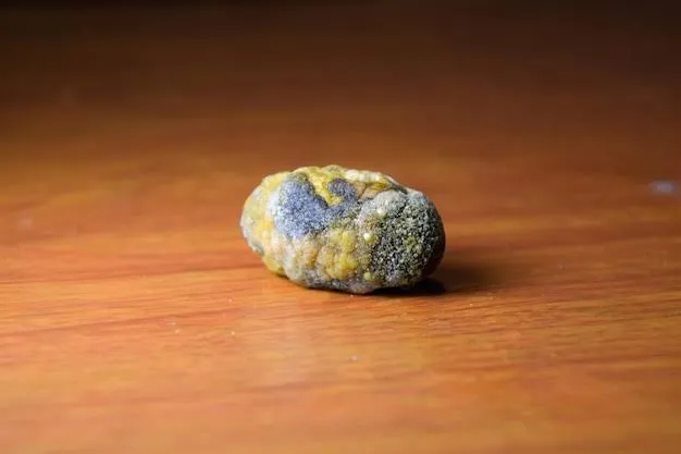
A patient has been suffering from gallbladder disease. A cholecystectomy has been performed and after evaluating the gallbladder sample of that Patient, the following stone is seen. Identify this stone?
Appeared in: NORCET 2 -2021 (Shift-2)
Explanation
- The stone in the image is a mixed gallstone, which is the most common type, accounting for about 80% of all gallstones.
- Its appearance is characteristic: a rough, mottled surface with yellowish areas (indicating cholesterol) and dark patches (indicating bile pigments and calcium salts).
- Mixed stones are composed of more than 50% cholesterol monohydrate, combined with an admixture of calcium salts, bile pigments, and fatty acids.
- Their formation is strongly associated with chronic cholecystitis (inflammation of the gallbladder).
Why Other Options Were Wrong
- Option A: Bile stones, also known as pigment stones, are primarily composed of calcium bilirubinate. They are typically small, brittle, and uniformly dark (either black or brown), which does not match the multi-colored, mottled appearance of the stone in the image.
- Option B: Oxalate stones are a common type of kidney stone (renal calculus), not a gallstone. They are formed in the urinary tract from calcium oxalate and are not found in the gallbladder.
- Option C: Uric acid stones are another type of kidney stone, not a gallstone. They form in acidic urine and are not related to the biliary system.
Related Visual

- Visual 1: Infographic: A comparative chart showing the three main types of gallstones (Cholesterol, Pigment, and Mixed) side-by-side, detailing their appearance, composition, and key characteristics.
- Visual 2: Diagram: A cross-section of a mixed gallstone, illustrating its laminated structure with alternating layers of yellow cholesterol and dark pigment/calcium.
- Visual 3: Illustration: The anatomy of the biliary system (liver, gallbladder, bile ducts) to clarify where gallstones form versus where kidney stones form.
Clinical Relevance
- Nursing practice connection: Safe nursing care depends on performing Identification of gallstone types based on macroscopic appearance in the correct sequence, documenting the action clearly, and monitoring for the expected response.
- Identifying the type of gallstone helps in understanding the underlying pathophysiology. Mixed and cholesterol stones are often linked to metabolic factors, while pigment stones are associated with conditions like hemolysis or infection.
- The risk factors for cholesterol and mixed stones are famously remembered by the '4 Fs': Female, Forty (age over 40), Fertile (multiparous), and Fat (obesity). This helps nurses in risk assessment and patient education.
- Post-cholecystectomy nursing care involves managing pain, monitoring for complications like infection or bile leakage, and providing dietary education (e.g., starting with a low-fat diet).
How to Approach the Question
- First, analyze the clinical context provided in the question stem. The key information is that the stone is from the 'gallbladder' following a 'cholecystectomy'.
- This immediately allows you to eliminate options that describe stones found in other organs. Both oxalate and uric acid stones are primarily kidney stones.
- Next, carefully examine the image of the stone. Note its key visual features: color (multiple colors - yellow and dark), texture (rough, mottled), and overall appearance (heterogeneous).
- Compare these visual features to the known characteristics of the remaining gallstone types (Bile/Pigment stone vs. Mixed stone).
- A pure bile/pigment stone is typically uniformly dark. A mixed stone is characteristically multi-colored and mottled, exactly like the one shown.
- Conclude that the stone's appearance perfectly matches the description of a mixed gallstone.
Concept Tested & Keywords
- Concept Tested: Identification of gallstone types based on macroscopic appearance.
- Stem keywords: gallbladder disease, cholecystectomy, gallbladder sample, stone
- Lead-in keywords: Identify
- Clinical cues: The location (gallbladder) is a critical clue to eliminate kidney stone types.
Question ID
QZdZATyK0z10YdD0TNCgqJ
Practise the full NORCET 2 -2021 (Shift-2)
Attempt every question from this paper in a timed mock, then review the full solution for each one.
More Systemic Pathology Questions
- The primary granuloma formed in tuberculosis is known as
- Which type of gall stone is related to hemolytic anemia?
- In HIV, the virus cannot be transmitted by?
- Type I hypersensitivity is mediated by?
- An example of type IV hypersensitivity is?
- Cystic fibrosis is an example of?
- Reed-Sternberg cells in nodes are found in?
- Hirschsprung disease is associated with a defect in which of the following developmental processes involving the enteric nervous system?
- A 36-year-old woman presents with secondary amenorrhea. Karyotyping shows 45,XO. What is the most likely diagnosis?
- What is the most common cause of gastric cancer?
More NORCET 2 -2021 (Shift-2) Questions
- Preferred route of tranexamic acid drug administration is?
- Parts of the fallopian tube, except?
- After thyroidectomy the patient complains for hoarseness of voice that occurs due to injury in?
- Highest source of vitamin-C?
- After the RTA, a patient was brought to the emergency department. After Massive blood transfusion, which electrolyte imbalance occurs in the patient?
- A patient suffering from shock is receiving normal saline (NS). What is the normal osmolarity of Na\ in NS?
- A patient is unconscious nurse is going to take the sterile urine sample for culture from the urinary catheter. What technique is to be used by the n…
- Covid face shield is sterilized by?
- Which reflex should check when death of the brain stem occurs, except?
- Which is an example of a tocolytic agent?