
GMCH Nursing Officer-2025
Medical Surgical Nursing
Medium
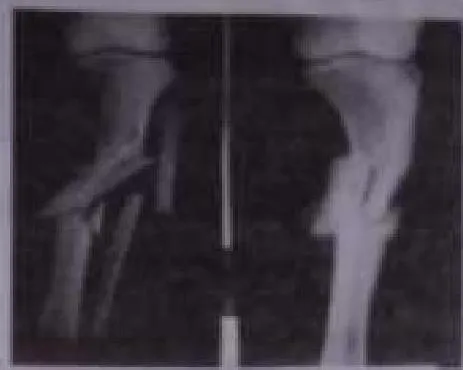
Identify the type of fracture seen in the given X-ray:
Appeared in: GMCH Nursing Officer-2025
Explanation
- A comminuted fracture is defined as a break or splinter of the bone into three or more pieces.
- The provided X-ray clearly shows the long bone is shattered into multiple fragments, which is the characteristic presentation of a comminuted fracture.
- This type of fracture typically results from high-energy trauma.
Why Other Options Were Wrong
- Option A: A transverse fracture is a straight break across the bone (at a right angle to the long axis). The X-ray does not show a single, clean horizontal break but rather multiple fragments.
- Option B: A greenstick fracture is an incomplete break, where the bone bends and cracks but does not separate completely. The image shows a complete fracture with multiple separate pieces.
- Option D: A spiral fracture has a fracture line that twists around the bone, caused by a rotational force. The fracture in the image is characterized by fragmentation, not a spiral pattern.
Related Visual

- Visual 1: Diagram - An illustration comparing different types of bone fractures (transverse, oblique, spiral, comminuted, greenstick) side-by-side to highlight their distinct patterns.
- Visual 2: X-ray Series - A collection of X-ray images, each showing a clear example of one of the major fracture types, allowing for direct comparison with the question's image.
Clinical Relevance
- Nursing practice connection: This is primarily an exam-oriented knowledge point with limited direct bedside application, so retain Identification of fracture types based on radiographic imaging as background academic context rather than a clinical decision trigger.
- Comminuted fractures are considered severe injuries due to the high energy required to cause them and the involvement of multiple bone fragments.
- These fractures often require surgical intervention, such as open reduction and internal fixation (ORIF), to realign the bone fragments and stabilize them with plates, screws, or rods.
- Nurses must perform meticulous neurovascular assessments (checking circulation, sensation, and movement) distal to the fracture site, as the sharp bone fragments can damage surrounding nerves and blood vessels.
How to Approach the Question
- First, carefully examine the provided X-ray image. Focus on the break in the bone.
- Ask yourself key questions about the fracture's appearance: Is the bone broken into two pieces or multiple fragments? Is the break line straight, angled, or spiral? Is the break complete or incomplete?
- In this image, observe that the bone is shattered into several pieces.
- Next, review the definitions of the fracture types listed in the options.
- Match your observation from the X-ray to the correct definition. The presence of multiple bone fragments directly corresponds to the definition of a comminuted fracture.
Concept Tested & Keywords
- Concept Tested: Identification of fracture types based on radiographic imaging.
- Stem keywords: fracture, X-ray
- Lead-in keywords: Identify
Question ID
Q1XiiL6hH66N9oFWBzNQfE
Practise the full GMCH Nursing Officer-2025
Attempt every question from this paper in a timed mock, then review the full solution for each one.
More Musculoskeletal System Questions
- Best way of healing a fracture is?
- The bandage that is applied over an amputated knee stump while in preparation for a prosthesis is usually a:
- Guerin sign seen in which diseases?
- Which of the following is inappropriate advice by a nurse caring for a patient with total hip replacement?
- The number of peshis in females:
- Paget's disease is related to which?
- Another name for osteitis deformans?
- Thomas splint is used to stabilize which bone?
- What is the cause of Osteomalacia in adults?
- Age-related disorder in which bone mass decreases and the likelihood of fractures increases.
More GMCH Nursing Officer-2025 Questions
- The burned area on the right arm of a patient received in emergency room appears red, has blisters, and is very painful. The nurse correctly identifi…
- Which of the following advice given by nurse to a postoperative patient of extracapsular cataract extraction and lens implantation is inappropriate?
- Nurse is giving care to a teenage girl with Mullerian agenesis on first postoperative day of vaginoplasty. Which of the following instruction by the…
- A patient (weight: 60 kg) admitted to critical care unit is to be administered with inj. methyl prednisolone 5.4 mg/kg/hour. The nurse dilutes 2 g me…
- Mrs. X is in active labor and is hyperventilating with a respiratory rate of 28. You are expecting that she will develop respiratory alkalosis. Which…
- Mrs. Y is a 24-year-old primigravida at 39 weeks gestation who presents to the labor and delivery room in active labor. On admission, the obstetricia…
- A woman who has been in labor for 36 hours is found to have a ruptured uterus. What is the most appropriate treatment in this scenario?
- Breast milk can be stored at room temperature safely for hours.
- The nurse identifies that the condition in which arterial blood gas analysis showing pH of 7 and plasma bicarbonate value of 19 mmol/L is .
- Mr. X is admitted with traumatic brain injury in critical care unit. On assessment, he opens eyes to pain, produces incomprehensible sound and locali…