
NORCET 2 -2021 (Shift-2)
Medical & Surgical Nursing
Medium
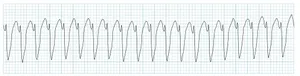
Identify the following ECG reading denoted in image?
Appeared in: NORCET 2 -2021 (Shift-2)
Explanation
- The ECG demonstrates a wide complex tachycardia, which is a key feature of rhythms originating in the ventricles.
- The rhythm is regular, with consistent R-R intervals, pointing towards a single ectopic focus.
- The heart rate is rapid, clearly over 100 beats per minute, meeting the definition of tachycardia.
- The QRS complexes are wide (greater than 0.12 seconds) and have a uniform (monomorphic) shape.
- P waves are not discernible, indicating the sinoatrial (SA) node is not in control of the ventricular rate.
- These findings collectively are the hallmark of monomorphic Ventricular Tachycardia.
Why Other Options Were Wrong
- Option A: Atrial Fibrillation is characterized by an 'irregularly irregular' rhythm and typically narrow QRS complexes. The ECG in the image shows a perfectly regular rhythm with wide QRS complexes.
- Option C: Ventricular Fibrillation is a chaotic, disorganized rhythm with no identifiable QRS complexes. The ECG in the image shows organized, distinct, and repeating QRS complexes.
- Option D: Atrial Tachycardia is a supraventricular rhythm, meaning it originates above the ventricles. Therefore, it typically produces narrow QRS complexes (less than 0.12 seconds). The ECG in the image clearly shows wide QRS complexes.
Related Visual

Clinical Relevance
- Nursing practice connection: This is primarily an exam-oriented knowledge point with limited direct bedside application, so retain ECG interpretation of life-threatening cardiac arrhythmias as background academic context rather than a clinical decision trigger.
- Ventricular Tachycardia is a medical emergency as it can severely decrease cardiac output, leading to hypotension, syncope, and shock.
- A critical nursing action is to immediately assess the patient's responsiveness and check for a pulse to determine if the VT is stable or unstable (pulseless).
- Sustained VT can quickly degenerate into Ventricular Fibrillation, which is a fatal arrhythmia if not treated with immediate defibrillation.
How to Approach the Question
- This is an image-based question requiring ECG interpretation. Use a systematic approach.
- Step 1: Analyze the Rate. Count the number of large squares between two R waves and divide 300 by that number. Here, it's roughly 1.5 large squares (300/1.5 = 200 bpm), which is a tachycardia.
- Step 2: Analyze the Rhythm. Check if the R-R intervals are regular or irregular. In the image, the intervals are consistent, so the rhythm is regular.
- Step 3: Analyze the P waves. Look for P waves before each QRS. In this strip, P waves are not visible.
- Step 4: Analyze the QRS duration. Measure the width of the QRS complex. Here, it is clearly wider than 3 small squares (0.12 seconds), making it a wide complex.
- Step 5: Synthesize the findings. A regular, rapid, wide-complex tachycardia with no visible P waves is the classic definition of Ventricular Tachycardia. This allows you to confidently select the correct option and rule out the distractors.
Concept Tested & Keywords
- Concept Tested: ECG interpretation of life-threatening cardiac arrhythmias.
- Stem keywords: ECG reading, image, identify
- Lead-in keywords: Identify
- Clinical cues: The visual data from the ECG strip is the primary clinical cue.
Question ID
Qcm8cdsFyFrV7NSqYAcML-
Reference Book
E6 Nursing Fundamentals Potter Perry 12e Part 4 p. 234-236
E6 Medicine Harrison 22e Part 1 p. 1996-1998
E6 Robert Boland, Marcia L. Verduin - Kaplan and Sadock's Comprehensive Text of Psychiatry-Wolters Kluwer Health (2024) (pp 1-16525 of 16525) p. 7321-7323
Practise the full NORCET 2 -2021 (Shift-2)
Attempt every question from this paper in a timed mock, then review the full solution for each one.
More Cardiovascular, Circulatory, and Hematologic Function Questions
- A patient is undergoing blood transfusion and suddenly shows complaints of transfusion reaction. What will be the priority nursing intervention?
- The following all are blood transfusion complications from; EXCEPT:
- Identify the use of following machine used in pneumatic compression device:
- In iron deficiency anemia, the fingernails become spoon shaped, this condition is referred to as:
- Which gauze cannula is commonly used for blood transfusion?
- If 'P' wave is absent in ECG rhythm, it is an indication of:
- Normal compression to ventilation ratio:
- Trans-illumination procedure is done with:
- A patient in the ward experiences sudden cardiac arrest. What is the most appropriate technique for opening the airway to facilitate endotracheal int…
- A patient experiences complications after a blood transfusion. Which of the following signs and symptoms is indicative of circulatory overload?
More NORCET 2 -2021 (Shift-2) Questions
- Preferred route of tranexamic acid drug administration is?
- Parts of the fallopian tube, except?
- After thyroidectomy the patient complains for hoarseness of voice that occurs due to injury in?
- Highest source of vitamin-C?
- After the RTA, a patient was brought to the emergency department. After Massive blood transfusion, which electrolyte imbalance occurs in the patient?
- A patient suffering from shock is receiving normal saline (NS). What is the normal osmolarity of Na\ in NS?
- A patient is unconscious nurse is going to take the sterile urine sample for culture from the urinary catheter. What technique is to be used by the n…
- Covid face shield is sterilized by?
- Which reflex should check when death of the brain stem occurs, except?
- Which is an example of a tocolytic agent?