
NORCET 5 Prelims - Sept 2023 (Shift-2)
Obstetrics & Gynaecology
Easy
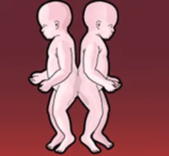
Find out the following image?
Appeared in: NORCET 5 Prelims - Sept 2023 (Shift-2)
Explanation
- The image shows two infants joined back-to-back at the buttocks and lower spine.
- This anatomical point of fusion is known as the sacrococcygeal region.
- The term 'Pygopagus' is derived from the Greek words 'pyge' (buttocks) and 'pagos' (fixed or joined), accurately describing the condition shown.
Why Other Options Were Wrong
- Option A: Ischiopagus twins are joined at the pelvis (ischium), not the buttocks. The image clearly shows fusion higher up, at the sacral region.
- Option B: Craniopagus refers to twins joined at the head (cranium). The image shows no fusion at the head.
- Option D: Omphalopagus twins are joined at the abdomen, usually near the navel. The image shows a posterior (back) fusion, not an anterior (front) abdominal fusion.
Related Visual

- Visual 1: Diagram - An illustration showing the four main types of conjoined twins (Thoracopagus, Omphalopagus, Pygopagus, Craniopagus) with labels indicating the point of fusion.
- Visual 2: Anatomical Drawing - A detailed medical illustration of Pygopagus twins, highlighting potentially shared structures like the lower spinal cord, rectum, and genitourinary system.
Clinical Relevance
- Nursing practice connection: This is primarily an exam-oriented knowledge point with limited direct bedside application, so retain Identification of types of conjoined twins as background academic context rather than a clinical decision trigger.
- The classification of conjoined twins is the first and most critical step in management, as it dictates the necessary diagnostic imaging (MRI, CT scans) to determine the extent of shared organs.
- For Pygopagus twins, nurses must be vigilant about potential shared anatomy of the gastrointestinal and genitourinary tracts, which can affect elimination and require specialized care for skin integrity and infection prevention.
- What if? If the twins were joined at the chest (Thoracopagus), the immediate concern would be a shared heart or major blood vessels, which has a much poorer prognosis and makes surgical separation significantly more complex and risky than for most Pygopagus cases.
How to Approach the Question
- First, carefully analyze the provided image to identify the key visual feature, which is the specific location where the twins are joined.
- Observe that the twins are fused back-to-back at the buttocks/lower spine area.
- Next, review the definitions of the terms provided in the options. The prefixes often give clues (e.g., 'cranio-' for head, 'omphalo-' for navel).
- Match your visual observation with the correct medical term. 'Pygopagus' refers to fusion at the buttocks.
- Eliminate the other options based on their definitions: Ischiopagus (pelvis), Craniopagus (head), and Omphalopagus (abdomen) do not match the image.
Concept Tested & Keywords
- Concept Tested: Identification of types of conjoined twins
- Stem keywords: conjoined twins, image
- Lead-in keywords: Find out
Question ID
QkEHN6VjyujTc-XDdeyVbq
Practise the full NORCET 5 Prelims - Sept 2023 (Shift-2)
Attempt every question from this paper in a timed mock, then review the full solution for each one.
More Multiple Pregnancy, Amniotic Fluid Disorders, Abnormalities of Placenta and Cord Questions
- Oligohydramnios is related with which of the following condition?
- Normal PH of amniotic fluid?
- A case of 35 weeks pregnancy with poly-hydramnios and marked respiratory distress is managed by?
- The functions of amniotic fluid are? a) Cushions the fetus b) Allows fetal movement c) Nutrient transport & x20; d) Prevents adhesions & x20;
- In which type of placental abnormality there is one or more lobes of placenta placed at varying distance from main placental margin?
- The water in the amniotic fluid is completely changed and replaced:
- What is normal amniotic fluid volume at term?
- What is the primary feature of oligohydramnios?
- A woman in labour with a twin pregnancy is being monitored. The first twin shows a low fetal heart rate. Suddenly the second twin's heart sounds beco…
- An ultrasound of a twin pregnancy reveals a single placenta and a thin dividing membrane between the two fetuses. This finding is characteristic of w…
More NORCET 5 Prelims - Sept 2023 (Shift-2) Questions
- A doctor ordered 0.45% normal saline with 50 ml/hr by microdrip set then what is drop per minute?
- In which of the following type of poisoning bluish line on gums seen?
- How many Anatomical regions are present in abdomen?
- Which is a preferred Foley catheter size used in adult female?
- A client was admitted with rib fractures and a Pneumothorax, which were sustained as a result of a motor vehicle accident. A chest tube was placed on…
- After a tonsillectomy, a child begins to vomit bright red blood. The nurse would take which initial action?
- A nurse is assessing a malnourished adolescent who has been consuming a vegan diet. For signs of which specific vitamin deficiency should the nurse a…
- A client is admitted to the emergency room with symptoms of delirium tremens. After admitting the client to a private room, the priority nursing inte…
- In which of the following consists with Charcot's triad, EXCEPT?
- The nurse is going to take perineal anus examination in a woman who had normally delivered a new born 2 days before. The nurse should place the woman…