
Combined AIIMS - 2020 (Jodhpur, Bhopal, Patna, Raipur)
Medical & Surgical Nursing
Easy
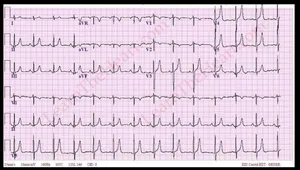
Diagnose the underlying medical disorder by ECG change in photograph:
Appeared in: Combined AIIMS - 2020 (Jodhpur, Bhopal, Patna, Raipur)
Explanation
- The ECG in the image shows tall, peaked, and narrow-based T-waves, which are the earliest and most common sign of hyperkalemia.
- This specific T-wave morphology, often described as 'tented', is due to altered ventricular repolarization caused by elevated extracellular potassium levels.
- Other subtle changes like a slightly widened QRS complex may also be present, indicating progression of the condition.
- These findings collectively point to a diagnosis of hyperkalemia.
Why Other Options Were Wrong
- Option A: Hypokalemia presents with ECG changes that are opposite to what is shown. It causes ST depression, T-wave flattening or inversion, and the appearance of a prominent U wave.
- Option B: Hypercalcemia is characterized by a shortened QT interval. The ECG in the image does not show a shortened QT interval; its most prominent feature is the abnormal T-wave morphology.
- Option C: Hypocalcemia leads to a prolonged QT interval due to a lengthened ST segment. The ECG shown does not demonstrate QT prolongation as the primary feature.
Related Visual

Clinical Relevance
- Nursing practice connection: This is primarily an exam-oriented knowledge point with limited direct bedside application, so retain ECG Interpretation in Electrolyte Imbalances as background academic context rather than a clinical decision trigger.
- Recognizing hyperkalemia on an ECG is a critical nursing skill because it is a medical emergency that can lead to life-threatening cardiac arrhythmias and cardiac arrest.
- A nurse observing these ECG changes must immediately notify the physician, anticipate orders for a stat serum potassium level, and prepare for emergency interventions.
- Emergency management includes stabilizing the cardiac membrane with intravenous calcium gluconate, shifting potassium into cells with insulin and dextrose, and removing potassium from the body using diuretics or potassium-binding resins (e.g., Kayexalate).
How to Approach the Question
- This is an image-based question that requires you to identify a classic ECG pattern.
- First, systematically analyze the ECG strip. Look at the P wave, PR interval, QRS complex, ST segment, and T wave in multiple leads.
- Identify the most prominent abnormality. In this ECG, the T-waves are strikingly tall and peaked ('tented'), especially in the precordial leads.
- Recall the characteristic ECG findings for the electrolyte imbalances listed in the options.
- Match the observed pattern (tall, peaked T-waves) with the correct diagnosis. This pattern is the hallmark of hyperkalemia.
- Rule out the other options by confirming their ECG patterns (e.g., hypokalemia causes flat T-waves and U-waves) do not match the image.
Concept Tested & Keywords
- Concept Tested: ECG Interpretation in Electrolyte Imbalances
- Stem keywords: ECG change, diagnose, medical disorder, photograph
- Lead-in keywords: Diagnose
- Clinical cues: The visual cue is the tall, peaked T-wave, which is a hallmark sign of hyperkalemia.
Question ID
Qt1BvVdE5JRokEyH3v7mLY
Reference Book
E6 Medicine Harrison 22e Part 1 p. 1915-1917
E6 Nelson Textbook of Pediatrics(2024) — Volume 1 p. 542-544
E6 Gynecology Williams p. 64-85
Practise the full Combined AIIMS - 2020 (Jodhpur, Bhopal, Patna, Raipur)
Attempt every question from this paper in a timed mock, then review the full solution for each one.
More Cardiovascular, Circulatory, and Hematologic Function Questions
- A patient is undergoing blood transfusion and suddenly shows complaints of transfusion reaction. What will be the priority nursing intervention?
- The following all are blood transfusion complications from; EXCEPT:
- Identify the use of following machine used in pneumatic compression device:
- In iron deficiency anemia, the fingernails become spoon shaped, this condition is referred to as:
- Which gauze cannula is commonly used for blood transfusion?
- If 'P' wave is absent in ECG rhythm, it is an indication of:
- Normal compression to ventilation ratio:
- Trans-illumination procedure is done with:
- A patient in the ward experiences sudden cardiac arrest. What is the most appropriate technique for opening the airway to facilitate endotracheal int…
- A patient experiences complications after a blood transfusion. Which of the following signs and symptoms is indicative of circulatory overload?
More Combined AIIMS - 2020 (Jodhpur, Bhopal, Patna, Raipur) Questions
- The position used for most of the gynecological procedures?
- Thyroid eye disease (TED) is also known as:
- How many items are included in the WHO surgical safety checklist: SITOSO?
- Which of the following code is associated with cardiac arrest during medical emergency?
- How is blood loss measured in Post-partum hemorrhage?
- The traction used for cervical spine injury:
- In operation theater during cauterization, the electric current passing from a single electrode is called:
- Tracheostomy is done in which tracheal ring?
- The instrument used for measuring fetal heart rate (FHR):