
INI-CET EXAM -2025
Medical & Surgical Nursing
Easy
A patient reports sudden onset of intense eye pain, headache, blurred vision, and colored halos. Examination reveals a mid-dilated fixed pupil, hazy cornea, and markedly raised intraocular pressure. What is the most probable diagnosis?
Appeared in: INI-CET EXAM -2025
Explanation
- The patient's constellation of symptoms—sudden severe eye pain, blurred vision with halos, and headache—is characteristic of a rapid spike in intraocular pressure (IOP).
- The physical examination findings of a mid-dilated fixed pupil, a hazy or steamy cornea, and markedly elevated IOP confirm the diagnosis.
- This clinical picture is the classic presentation of Acute Primary Angle Closure Glaucoma (PACG), where the drainage angle for aqueous humor is abruptly blocked.
- The condition is an ophthalmologic emergency because the high pressure can quickly cause irreversible damage to the optic nerve, leading to permanent vision loss.
Why Other Options Were Wrong
- Option A: While anterior uveitis can cause secondary angle closure, the primary presentation typically involves signs of intraocular inflammation like keratic precipitates and cells in the anterior chamber, which are not mentioned. The scenario describes a classic primary angle closure event.
- Option B: Acute endophthalmitis is a severe intraocular infection. While it causes pain and vision loss, it is typically associated with a history of recent eye surgery or trauma and often presents with a hypopyon (pus in the anterior chamber), none of which are described.
- Option C: Orbital cellulitis is an infection of the fat and muscles surrounding the eye. Its hallmark signs are proptosis (bulging eye), pain with eye movements, and restricted ocular motility. The patient's signs are confined to the globe itself (cornea, pupil, IOP).
Related Visual
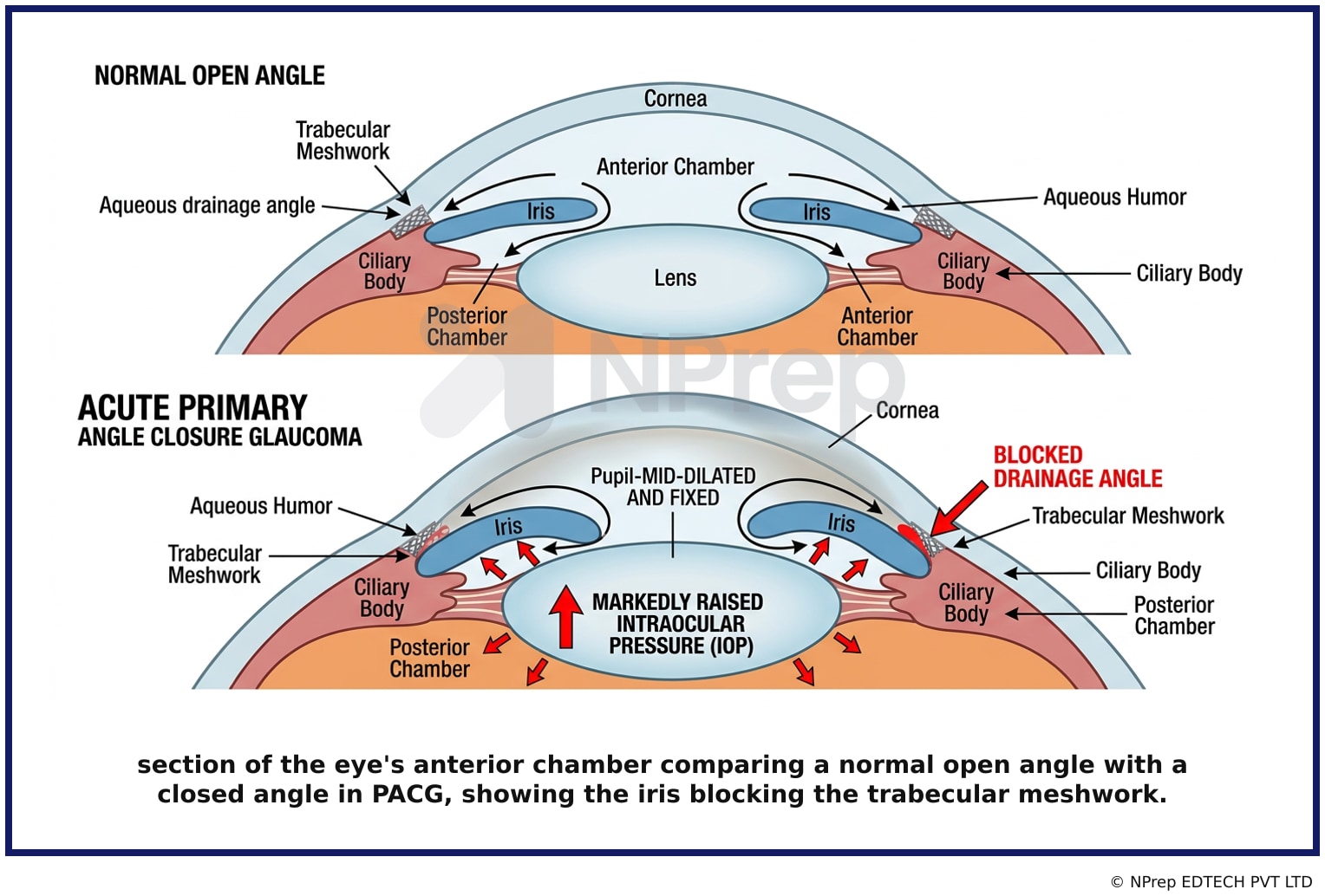
- Visual 1: Diagram - A cross-section of the eye's anterior chamber comparing a normal open angle with a closed angle in PACG, showing the iris blocking the trabecular meshwork.
- Visual 2: Clinical Image - A photograph of an eye during an acute angle closure attack, highlighting the hazy/cloudy cornea and a mid-dilated, oval-shaped pupil.
Clinical Relevance
- Nursing practice connection: Use the key finding related to Diagnosis of Acute Angle Closure Glaucoma to guide bedside assessment, documentation, and the next nursing action.
- Acute Primary Angle Closure Glaucoma is a true ophthalmologic emergency. Delay in treatment can lead to permanent optic nerve damage and irreversible blindness within hours to days.
- Nurses in any setting (ER, clinic, general ward) must be able to recognize this classic symptom cluster. Prompt identification and immediate referral to an ophthalmologist are critical nursing actions.
- What if the patient only reported blurred vision and halos without severe pain? This could indicate subacute or intermittent angle closure, which is less dramatic but still requires urgent ophthalmologic evaluation to prevent a full-blown acute attack.
How to Approach the Question
- First, analyze the patient's symptoms: identify them as acute, severe, and localized to the eye with systemic components (headache). Note keywords like 'sudden,' 'intense,' 'halos.'
- Next, correlate the symptoms with the objective examination findings: a mid-dilated fixed pupil, hazy cornea, and high IOP.
- Recognize that this specific combination of signs and symptoms forms a classic clinical triad.
- Evaluate the options. The correct diagnosis must account for all the key features presented.
- Eliminate distractors by identifying their typical presentations. Orbital cellulitis involves the orbit, not just the globe. Endophthalmitis involves infection/pus. Secondary glaucoma from uveitis would have primary inflammatory signs.
- Select the diagnosis, Acute Primary Angle Closure Glaucoma, that perfectly matches the entire clinical picture described.
Concept Tested & Keywords
- Concept Tested: Diagnosis of Acute Angle Closure Glaucoma
- Stem keywords: sudden onset, intense eye pain, headache, blurred vision, colored halos, mid-dilated fixed pupil, hazy cornea, raised intraocular pressure
- Lead-in keywords: most probable diagnosis
- Clinical cues: The combination of a fixed mid-dilated pupil, hazy cornea, and sudden severe pain is a classic triad for acute angle closure glaucoma.
Question ID
QArYvtruaxDjOLA6FQobU4
Practise the full INI-CET EXAM -2025
Attempt every question from this paper in a timed mock, then review the full solution for each one.
More Sensorineural Function Questions
- Which ophthalmic diagnostic test measures the Intraocular pressure?
- Identify the following instrument in image:
- What sound is heard when percussing a distended bladder?
- Which test would be done to check balance in a patient with ataxia?
- Identify the condition:
- In the emergency you have 4 patients. By prioritizing your management plan, which patient's problem will you address first?:
- All of the following instructions are given to a patient before computed tomography (CT) scan, except?
- The nursing care of a patient with dengue fever includes all of the following, except?
- Which of the following equipment is used for the examination of the ear?
- An aged client with diabetes mellitus complaining of painless peripheral loss of vision is at risk for?
More INI-CET EXAM -2025 Questions
- The highlighted structure in the provided image articulates with which bone?& x20;
- Through which of the following foramina does the nerve supplying the muscles of mastication pass?& x20;
- In the given image, which region is correctly paired with its corresponding brain structure? • Middle cerebellar peduncle • Fasciculus cuneatus • Sup…
- Which of the following muscles is NOT typically associated with dysphagia or dysarthria?
- A muscle of mastication involved in opening the mouth exhibits the following characteristics. Identify the incorrect statement about it:
- In the illustrated image, which of the following structures is NOT associated with the area marked by the arrow?& x20;
- Match the following embryonic aortic arches with their respective adult derivatives: A. 1st Arch 1. Maxillary artery& x20; B. 2nd Arch 2. Common caro…
- Which of the following histology image is not an endocrine structure?& x20;
- Marked structure separates which of the following:
- Which of the following bones of the skull gives passage to a nerve arising from the brain & x20;