
DSSSB 13 August 2024
Medical Surgical Nursing
Medium
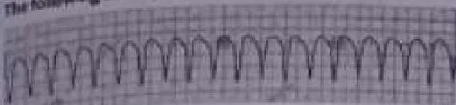
The following ECG represents:
Appeared in: DSSSB 13 August 2024
Explanation
- The ECG displays a heart rate of approximately 150-250 beats per minute, which is a tachycardia.
- The rhythm is regular, as indicated by the consistent distance between the QRS complexes.
- The QRS complexes are wide (greater than 0.12 seconds) and uniform in shape (monomorphic), which is a hallmark of an impulse originating in the ventricles.
- There are no visible P waves preceding the QRS complexes, indicating the rhythm is not originating from the sinoatrial node.
- This combination of a fast, regular, wide-complex tachycardia is the classic presentation of Ventricular Tachycardia (VT).
Why Other Options Were Wrong
- Option A: Atrial fibrillation is characterized by an 'irregularly irregular' rhythm and typically narrow QRS complexes. The ECG in the image shows a regular rhythm with wide complexes.
- Option B: Atrial flutter is identified by its characteristic 'sawtooth' flutter waves and usually has narrow QRS complexes. The ECG in the image lacks sawtooth waves and has wide QRS complexes.
- Option C: Ventricular fibrillation is a chaotic, disorganized rhythm with no identifiable QRS complexes, appearing as a quivering line. The ECG in the image shows organized, regular, and distinct QRS complexes.
Related Visual

Clinical Relevance
- Nursing practice connection: This is primarily an exam-oriented knowledge point with limited direct bedside application, so retain ECG interpretation of life-threatening cardiac arrhythmias as background academic context rather than a clinical decision trigger.
- Recognizing Ventricular Tachycardia is a critical skill for nurses, as it is a medical emergency that can quickly lead to cardiac arrest.
- The first nursing action upon seeing this rhythm on a monitor is to assess the patient for a pulse and level of consciousness. The treatment depends on whether the patient is stable (has a pulse and is conscious) or unstable (pulseless or unconscious).
- What if? If the patient with this rhythm was found to be pulseless, the situation would be treated as a cardiac arrest. The nurse should immediately call for help, start high-quality CPR, and prepare for defibrillation, as pulseless VT is a shockable rhythm.
How to Approach the Question
- First, analyze the image provided, which is an ECG rhythm strip.
- Systematically evaluate the ECG using a standard method. A common approach is the 5-step method: Rate, Rhythm, P wave, PR interval, and QRS duration.
- Step 1: Determine the rate. It is clearly fast (tachycardia).
- Step 2: Assess the rhythm. The distance between the tall complexes (R-R interval) is regular.
- Step 3: Look for P waves. No clear P waves are visible.
- Step 4: Measure the QRS duration. The complexes are very wide, clearly greater than 3 small squares (0.12 seconds).
Concept Tested & Keywords
- Concept Tested: ECG interpretation of life-threatening cardiac arrhythmias.
- Stem keywords: ECG, arrhythmia, electrocardiogram
- Lead-in keywords: represents
- Clinical cues: The visual evidence of a wide-complex tachycardia on the ECG strip is the primary cue.
Question ID
Q9DVzpUxBY7_woh0YXIS7l
Reference Book
E6 Pharmacology Nursing Lilley 11e Part 2 p. 86-88
E6 Medicine Harrison 22e Part 1 p. 1996-1998
Practise the full DSSSB 13 August 2024
Attempt every question from this paper in a timed mock, then review the full solution for each one.
More Cardiovascular System Questions
- What is pulse deficit?
- Difference between apical pulse and radial pulse is called?
- A 40 year old patient is admitted with crushing chest pain and an Electrocardiogram (ECG) showing Acute Myocardial Infarction. He is pale, has Blood…
- "Clubbing of the Nails" finding is found in which of the following disease conditions?
- Which of the following tests need to be performed before radial artery cannulation to evaluate radial and ulnar arterial patency?
- Which among the following is the most common cause of acute infective endocarditis?
- Levine's sign is usually associated with:
- In case of ventricular fibrillation, a nurse must prepare for the first step of treatment?
- A nurse is reviewing an ECG that shows ventricular tachycardia. Which of the following characteristics would be most evident on the ECG?
- The mula (adhishtana) of siraas is:
More DSSSB 13 August 2024 Questions
- Which of the following is NOT a component of mental status examination?
- Which therapy is being shown in the given image?
- Which of the following correctly describes esotropia?
- The tablet Arkaminis used in the management of:
- A patient was brought to the emergency department and a blood sample was sent for serum electrolyte level. The report shows that the serum potassium…
- The concept of therapeutic community was first developed by:
- The superego functions based on:
- Reed-Sternberg cells are distinctive, giant cells have light microscopy in biopsies from individuals with the following diseases?
- Which of the following intakes is usually associated with "Flashback phenomenon"?
- Which of the following drugs is commonly used to treat vertigo?