
AIIMS Manglagiri NO - 2019
Medical & Surgical Nursing
Easy
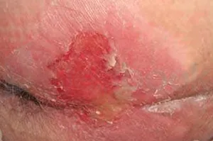
Identify the stage of pressure ulcer
Appeared in: AIIMS Manglagiri NO - 2019
Explanation
- The image displays a shallow, open ulcer with a red-pink, moist wound bed, which is a classic presentation of a Stage 2 pressure ulcer.
- This stage involves partial-thickness loss of the dermis, meaning the upper layers of the skin are gone, but deeper tissues like fat, muscle, or bone are not exposed.
- The absence of slough (yellow or tan dead tissue), eschar (black or brown necrotic tissue), and visible subcutaneous fat confirms that the ulcer has not progressed to Stage 3 or 4.
Why Other Options Were Wrong
- Option A: Stage 1 involves intact skin with non-blanchable redness. The image clearly shows broken skin, which rules out Stage 1.
- Option C: Stage 3 is defined by full-thickness skin loss where subcutaneous fat is visible. The ulcer in the image is shallow and does not show any adipose (fat) tissue.
- Option D: Stage 4 involves full-thickness skin and tissue loss with exposed muscle, tendon, or bone. The wound in the image is superficial and does not extend to these deeper structures.
Related Visual

Clinical Relevance
- Nursing practice connection: This is primarily an exam-oriented knowledge point with limited direct bedside application, so retain Staging of Pressure Ulcers as background academic context rather than a clinical decision trigger.
- Accurate staging of pressure ulcers is a critical nursing skill that directly impacts the plan of care, including dressing selection, pressure redistribution strategies, and nutritional support.
- Failure to correctly identify the stage can lead to improper treatment, delayed healing, and progression of the wound to a more severe stage, increasing the risk of infection and other complications.
- What if? If the wound bed in the image was covered by more than 50% yellow slough, it would be classified as an 'Unstageable Pressure Injury' because the slough obscures the true depth of the wound. The wound would need to be debrided before it could be accurately staged.
How to Approach the Question
- First, carefully examine the image of the wound. Note its key characteristics: depth, color of the wound bed, and the types of tissue visible.
- Observe that the skin is broken, forming a shallow crater. This immediately rules out Stage 1, which involves intact skin.
- Identify the tissue in the wound bed. It appears red and pink, which is the dermis. Note the absence of deeper tissues like yellow fat (subcutaneous), muscle, or bone.
- Recall the definitions for each pressure ulcer stage. A shallow ulcer with exposed dermis and no visible subcutaneous tissue matches the definition of a Stage 2 pressure ulcer.
- Compare your observation with the definitions for Stage 3 (visible fat) and Stage 4 (visible bone/muscle) to confirm they are incorrect.
- Select the option that corresponds to your conclusion.
Concept Tested & Keywords
- Concept Tested: Staging of Pressure Ulcers
- Stem keywords: pressure ulcer, stage
- Lead-in keywords: Identify
- Clinical cues: Image shows an open wound with a red-pink base.
Question ID
QFlbR7CQfJPd0dCe9zoz0f
Reference Book
E6 Nursing Fundamentals Potter Perry 12e Part 6 p. 69-71
E6 Nursing Fundamentals Taylor pp. 560-562, 581-583
Practise the full AIIMS Manglagiri NO - 2019
Attempt every question from this paper in a timed mock, then review the full solution for each one.
More Integumentary Function Questions
- This type of dressing is known as:
- The following picture shows is bedsore present in bedridden patient. Which grade of bedsore is present?
- In second degree bedsore, there is an involvement of:
- What is the bath used to remove the irritation of certain types of skin diseases and provide soothing effect?
- A female patient has come with facial burn injury into the emergency department in conscious condition. What will be the treatment for this patient?
- A patient comes into the emergency with a burn injury of 40% then what is first nursing intervention?
- A nurse caring a bedridden patient with a pressure ulcer, 2 hourly position changes and massaging & rubbing the back but pressure sore does not heal.…
- Second degree burn considered?
- A 23 year old man comes to the emergency department with thermal burns. In this case according to rule of nine which part of the body burned maximum?
- Identify the condition shown in image:
More AIIMS Manglagiri NO - 2019 Questions
- Graves' Disease is:-
- Braxton-Hicks are:-
- A function of nursing management is:-
- Calculate the sampling interval according to systematic sampling technique when your sample size is 200 and your accessible population is 40,000?
- Following are the dimensions of health except:-
- Basic concepts of nursing informatics:-
- A feeding problem among infants:-
- Malpractice is an example of:
- Dystocia is:-
- The third stage of labour comprises:-