
NORCET -4 , 2023
Medical & Surgical Nursing
Medium
Identify the following radiological finding which is denoting in image?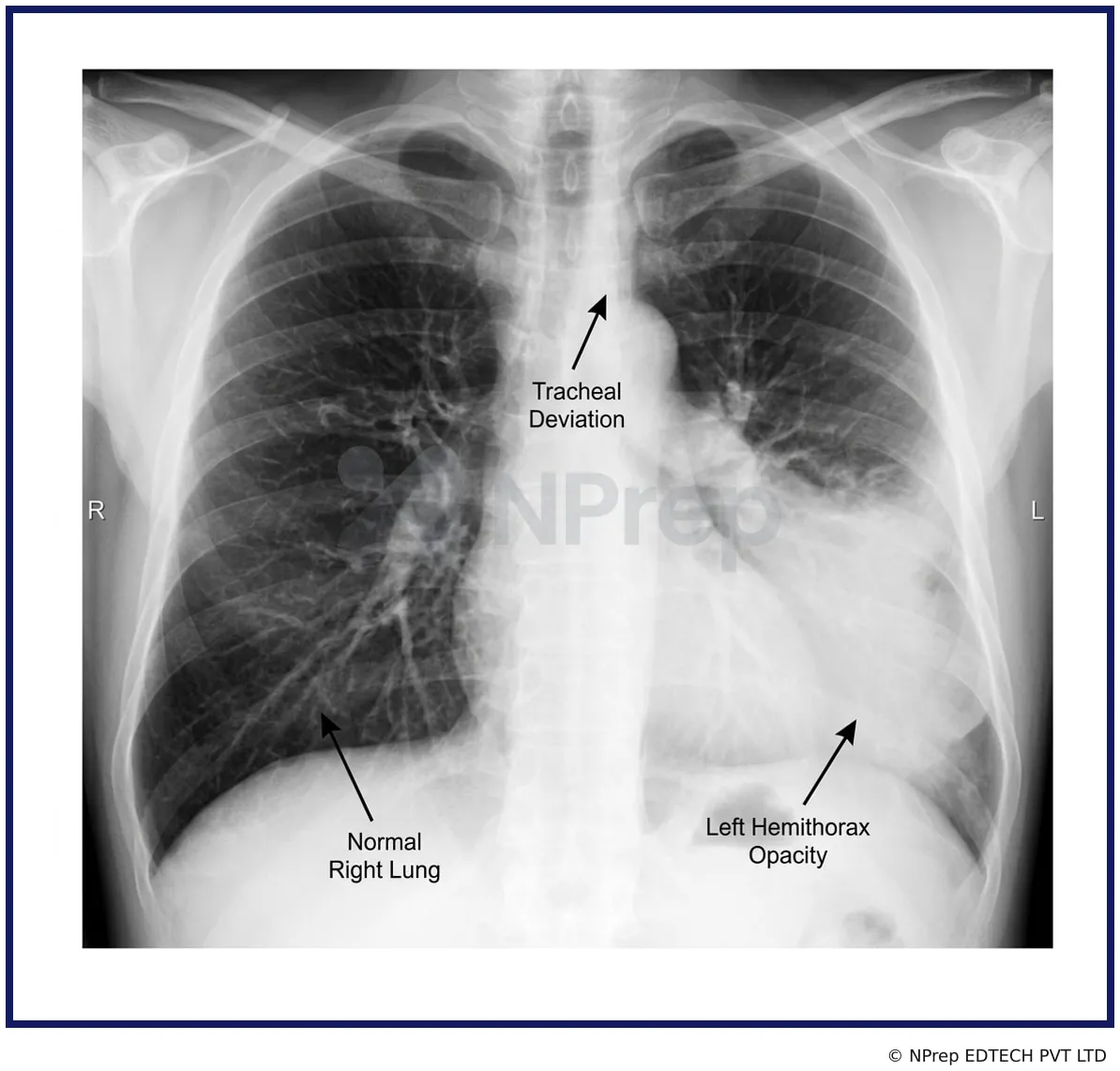
Appeared in: NORCET -4 , 2023
Explanation
- The correct diagnosis is pleural effusion, which is the accumulation of fluid in the pleural space.
- The chest X-ray shows a large, dense, homogeneous opacity in the left hemithorax, which is a classic sign of fluid.
- A key diagnostic feature in this image is the contralateral (opposite side) shift of the trachea and mediastinum to the right. This indicates that the pressure from the massive amount of fluid on the left is pushing these structures away.
- The costophrenic angle and the outline of the left hemidiaphragm are completely obscured by the opacity, further confirming the presence of fluid.
Why Other Options Were Wrong
- Option A: COPD (Chronic Obstructive Pulmonary Disease) is characterized by air trapping, not fluid accumulation. Its X-ray findings are the opposite of what is seen here.
- Option C: A tension pneumothorax involves air, not fluid, in the pleural space. This would appear as a hyperlucent (dark) area, not a dense white opacity.
- Option D: ARDS (Acute Respiratory Distress Syndrome) typically causes diffuse, bilateral (affecting both lungs) opacities, often described as a 'white-out'. The finding in the image is clearly unilateral (affecting only one side).
Related Visual

Clinical Relevance
- Nursing practice connection: This is primarily an exam-oriented knowledge point with limited direct bedside application, so retain Interpretation of chest X-ray for common respiratory conditions as background academic context rather than a clinical decision trigger.
- Recognizing a massive pleural effusion on a chest X-ray is a critical skill for nurses, as it indicates a life-threatening condition requiring urgent intervention.
- A massive effusion can cause severe respiratory distress and hemodynamic instability (e.g., low blood pressure, high heart rate) due to compression of the lung and heart.
- Nursing priorities include continuous monitoring of vital signs and oxygen saturation, administering supplemental oxygen, positioning the patient for comfort (often upright), and preparing for an emergency thoracentesis (needle drainage) or chest tube insertion.
How to Approach the Question
- First, systematically analyze the chest X-ray. Check the 'ABCDEs': Airway (trachea position), Breathing (lung fields, pleura), Cardiac (heart silhouette), Diaphragm, and Everything else (bones, soft tissues).
- Identify the primary abnormality. In this image, it's the large white opacity on the left.
- Note any secondary signs. The most striking secondary sign is the tracheal deviation to the right.
- Correlate the primary abnormality with the secondary signs. A large opacity (mass/fluid) that pushes the trachea away indicates high pressure on the affected side.
- Evaluate the given options based on this analysis. A pleural effusion is fluid. A massive effusion creates high pressure. This matches the findings.
- Rule out the other options by recalling their classic X-ray appearances: COPD (hyperinflation), tension pneumothorax (hyperlucency/air), and ARDS (bilateral opacities).
Concept Tested & Keywords
- Concept Tested: Interpretation of chest X-ray for common respiratory conditions.
- Stem keywords: radiological finding, chest X-ray, image
- Lead-in keywords: Identify
- Clinical cues: Left hemithorax opacity
- Clinical cues: Tracheal deviation to the right
Question ID
QZQo5CjkbNw0ChEP5d-NmK
Reference Book
E6 Nelson Textbook of Pediatrics(2024) — Volume 2 p. 444-446
Practise the full NORCET -4 , 2023
Attempt every question from this paper in a timed mock, then review the full solution for each one.
More Gas Exchange and Respiratory Function Questions
- Which is the most relevant knowledge about oxygen administration to a client with COPD?
- Patient in emergency department diagnosed to have fluid and air present in lung. What is the above-mentioned condition known as?
- Patient is suffering from dyspnea. Which position will you provide for patient to relieve?
- Which of the following is not involved in chest physiotherapy?
- Respiration which is abnormally shallow for two or three breaths followed by irregular period of apnea is termed:
- Identify the following instrument which is used in diagnostic procedures:
- Identify the instrument shown in the following image:
- Identify the tube in the following image:
- A patient suddenly develops respiratory difficulties such as dyspnea, what is the first priority of the nursing officer?
- Which of the following method is used for high flow oxygen administration:
More NORCET -4 , 2023 Questions
- A pregnant client comes to the office for her first prenatal examination at 10 weeks. She has been pregnant twice before, the first delivery produced…
- A client is admitted to the hospital for an induction of labor owing to a gestation of 42 weeks confirmed by dates and ultrasound. When she is dilate…
- Which of the following step is not included in surgical safety checklist?
- All of the following are correct about WHO 5 hand hygiene movement, EXCEPT?
- After liver biopsy, the patient should be placed in?
- What is the best position to keep a 12-year-old child after a lumbar puncture to assess CSF?
- When teaching a mother of a 4-month-old with diarrhoea about the importance of preventing dehydration, the nurse would inform the mother about the im…
- Prior to administration of antibiotics there is need to sensitivity test. Which of the following antibiotic is not required sensitivity test?
- A client with a cervical spine fracture at C, level has just arrived in the emergency room. The primary nursing intervention would be?
- A client is being discharged with albuterol and beclomethasone dipropionate to be administered via inhalation three times a day and at bedtime. Clien…