
NORCET -4 , 2023
Medical & Surgical Nursing
Easy
Identify the following ECG finding which is denoting in photograph?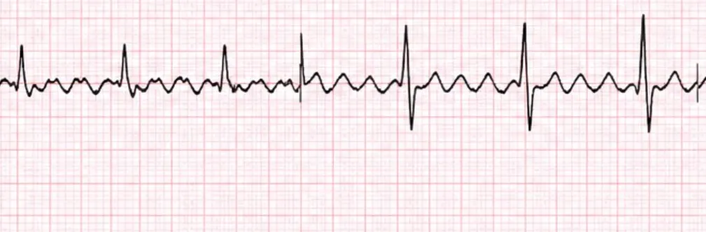
Appeared in: NORCET -4 , 2023
Explanation
- The ECG demonstrates classic 'sawtooth' flutter waves between the QRS complexes, which is the pathognomonic sign of Atrial Flutter.
- This rhythm originates from a large re-entrant circuit within the atria, leading to rapid but organized atrial depolarization at a rate of 250-350 bpm.
- The ventricular response is typically regular because the AV node conducts these impulses at a fixed ratio (e.g., 2:1, 3:1, or 4:1), as seen in the image.
- The QRS complexes are narrow, confirming that the origin of the rhythm is supraventricular (above the ventricles).
Why Other Options Were Wrong
- Option A: Atrial fibrillation is characterized by an 'irregularly irregular' rhythm and a chaotic baseline with no discernible P waves (fibrillatory waves), which is different from the regular rhythm and organized sawtooth waves seen in the image.
- Option B: Ventricular tachycardia originates in the ventricles, resulting in wide QRS complexes (greater than 0.12 seconds). The ECG in the image shows narrow QRS complexes, ruling out a ventricular origin.
- Option D: SVT (Supraventricular Tachycardia) is a broad term. While atrial flutter is a type of SVT, this option usually refers to other forms like AVNRT, which present as a rapid, regular rhythm but lack the distinct sawtooth pattern. 'Atrial flutter' is the most specific and accurate diagnosis.
Related Visual

Clinical Relevance
- Nursing practice connection: This is primarily an exam-oriented knowledge point with limited direct bedside application, so retain ECG interpretation of supraventricular tachyarrhythmias as background academic context rather than a clinical decision trigger.
- Like atrial fibrillation, atrial flutter significantly increases the risk of thromboembolism and stroke due to blood stasis in the non-contracting atria. Patients often require anticoagulation based on their CHA2DS2-VASc score.
- The primary goals of management are to control the ventricular rate (rate control), restore sinus rhythm (rhythm control), and prevent stroke.
- Achieving rate control in atrial flutter can sometimes be more difficult than in atrial fibrillation because of the organized nature of the atrial impulses.
How to Approach the Question
- First, analyze the rhythm by looking at the R-R intervals. In this ECG, they are regular.
- Next, examine the baseline for atrial activity. Instead of normal P waves, identify the characteristic 'sawtooth' pattern.
- Measure the QRS duration. Note that the QRS complexes are narrow, which points to a supraventricular origin.
- Based on the combination of a regular rhythm, narrow QRS, and sawtooth flutter waves, conclude the diagnosis is Atrial Flutter.
- Systematically rule out the other options: Atrial Fibrillation (irregularly irregular), Ventricular Tachycardia (wide QRS), and generic SVT (lacks sawtooth pattern).
Concept Tested & Keywords
- Concept Tested: ECG interpretation of supraventricular tachyarrhythmias.
- Stem keywords: ECG finding, photograph, arrhythmia
- Lead-in keywords: Identify
- Clinical cues: The presence of a sawtooth pattern on the ECG is a key diagnostic clue.
Question ID
Qsf2M9LK31kVqlqZFKEdxt
Reference Book
E6 Medicine Harrison 22e Part 1 p. 1984-1986
E6 Nelson Textbook of Pediatrics(2024) — Volume 2 p. 641-643
Practise the full NORCET -4 , 2023
Attempt every question from this paper in a timed mock, then review the full solution for each one.
More Cardiovascular, Circulatory, and Hematologic Function Questions
- A patient is undergoing blood transfusion and suddenly shows complaints of transfusion reaction. What will be the priority nursing intervention?
- The following all are blood transfusion complications from; EXCEPT:
- Identify the use of following machine used in pneumatic compression device:
- In iron deficiency anemia, the fingernails become spoon shaped, this condition is referred to as:
- Which gauze cannula is commonly used for blood transfusion?
- If 'P' wave is absent in ECG rhythm, it is an indication of:
- Normal compression to ventilation ratio:
- Trans-illumination procedure is done with:
- A patient in the ward experiences sudden cardiac arrest. What is the most appropriate technique for opening the airway to facilitate endotracheal int…
- A patient experiences complications after a blood transfusion. Which of the following signs and symptoms is indicative of circulatory overload?
More NORCET -4 , 2023 Questions
- A pregnant client comes to the office for her first prenatal examination at 10 weeks. She has been pregnant twice before, the first delivery produced…
- A client is admitted to the hospital for an induction of labor owing to a gestation of 42 weeks confirmed by dates and ultrasound. When she is dilate…
- Which of the following step is not included in surgical safety checklist?
- All of the following are correct about WHO 5 hand hygiene movement, EXCEPT?
- After liver biopsy, the patient should be placed in?
- What is the best position to keep a 12-year-old child after a lumbar puncture to assess CSF?
- When teaching a mother of a 4-month-old with diarrhoea about the importance of preventing dehydration, the nurse would inform the mother about the im…
- Prior to administration of antibiotics there is need to sensitivity test. Which of the following antibiotic is not required sensitivity test?
- A client with a cervical spine fracture at C, level has just arrived in the emergency room. The primary nursing intervention would be?
- A client is being discharged with albuterol and beclomethasone dipropionate to be administered via inhalation three times a day and at bedtime. Clien…