
NORCET 2 - 2021 (shift-1)
Medical & Surgical Nursing
Medium
Identify the following condition in the X-ray image.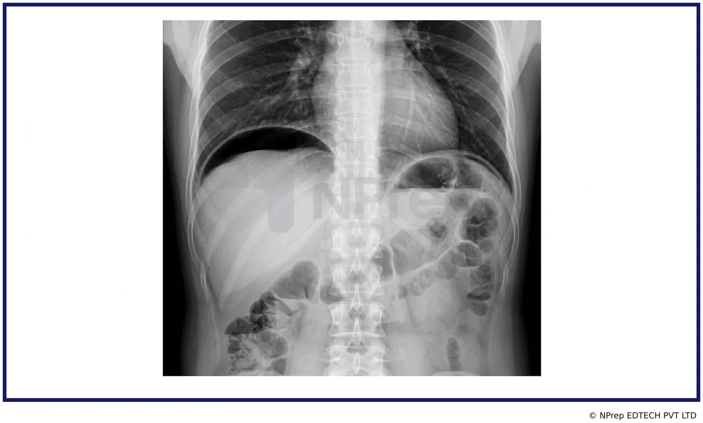
Appeared in: NORCET 2 - 2021 (shift-1)
Explanation
- The X-ray clearly displays a large, crescent-shaped lucency (dark area) between the liver and the right hemidiaphragm.
- This finding is the classic radiological sign of pneumoperitoneum, which is free air in the abdominal (peritoneal) cavity.
- Pneumoperitoneum is most often caused by the rupture or perforation of a hollow organ, such as the stomach or intestine.
- The leakage of bowel contents and air into the abdomen causes peritonitis, a serious and painful inflammation that requires urgent surgical intervention.
Why Other Options Were Wrong
- Option A: A right-sided pneumothorax involves air in the pleural space (in the chest cavity, surrounding the lung), not under the diaphragm. The X-ray would show the edge of the collapsed lung and an absence of lung markings in the periphery of the chest.
- Option C: Small intestinal obstruction (SAIO) typically presents with multiple dilated loops of the small bowel (appearing like a 'stack of coins') and air-fluid levels on an abdominal X-ray. While some bowel gas is visible, the dominant finding is free air, not obstruction.
- Option D: Upward protrusion of the diaphragm (eventration) would show an abnormally high position of the diaphragm itself, but there would not be a separate, distinct crescent of air between the diaphragm and the liver as seen in this image.
Related Visual

Clinical Relevance
- Nursing practice connection: This is primarily an exam-oriented knowledge point with limited direct bedside application, so retain Radiological interpretation of an erect chest X-ray for identifying pneumoperitoneum as background academic context rather than a clinical decision trigger.
- Pneumoperitoneum due to a perforated viscus is a surgical emergency. A nurse's prompt recognition and reporting of associated signs (e.g., sudden, severe abdominal pain, board-like rigidity) are critical for patient survival.
- Immediate nursing interventions include keeping the patient NPO (nothing by mouth), establishing IV access for fluids and antibiotics, preparing for an NG tube insertion to decompress the stomach, and continuous monitoring of vital signs for signs of septic shock.
- What if? If the patient was unable to sit or stand for the X-ray, a left lateral decubitus view would be ordered. In this position, free air would collect over the lateral aspect of the liver, appearing as a lucency between the liver and the abdominal wall.
How to Approach the Question
- First, systematically analyze the X-ray image. Look at the lungs, heart, bones, and the areas above and below the diaphragm.
- Identify the most striking abnormality. In this case, it is the large, dark, crescent-shaped area under the right side of the diaphragm.
- Recognize this specific sign. The crescent of air under the diaphragm is the hallmark of pneumoperitoneum.
- Consider the clinical implication of this sign. Pneumoperitoneum most commonly indicates a perforated hollow organ.
- Evaluate the given options. 'Perforated peritonitis' is the clinical condition directly associated with the radiological sign of pneumoperitoneum.
- Mentally exclude the other options by recalling their typical X-ray appearances, which do not match the image.
Concept Tested & Keywords
- Concept Tested: Radiological interpretation of an erect chest X-ray for identifying pneumoperitoneum.
- Stem keywords: X-ray image, condition
- Lead-in keywords: Identify
- Clinical cues: Large crescent of air under the right hemidiaphragm.
Question ID
Q_J9mG8B342h2FBMBMb7D6
Reference Book
E6 Nelson Textbook of Pediatrics(2024) — Volume 2 p. 492-494
E6 Nelson Textbook of Pediatrics(2024) — Volume 1 p. 1106-1108
Practise the full NORCET 2 - 2021 (shift-1)
Attempt every question from this paper in a timed mock, then review the full solution for each one.
More Digestive and Gastrointestinal Function Questions
- Which condition is defined as the inflammation of the peritoeum?
- Which types of stools are seen in Gastric bleeding?
- A patient comes to the ED with 105°F fever and hypertension and bradycardia also having vomiting and diarrhea past one day and he also has loosening…
- A patient complains of right abdominal tenderness, pain at the right lower quadrants region, nausea, vomiting and too much restlessness. In X-ray fil…
- A postoperative patient requires which nutrient after ileostomy?
- Which of the following clinical features are seen in intestinal obstruction, except?
- A patient comes in the ED with hematemesis and mild splenomegaly. What would be the possible cause?
- Ranson score is used in which clinical finding?
- In which of the following consists with Charcot's triad, EXCEPT?
- Following a gastric resection, which of the following actions would the nurse reinforce with the client in order to alleviate the distress from dumpi…
More NORCET 2 - 2021 (shift-1) Questions
- Which type of bandage used in ankle injury?
- A nurse knows that under the National Family Health Program, the duration of Copper T 380A is?
- Which diet should be avoided in acute renal failure disease?
- What was the first indigenous vaccine in India?
- Which bone is known as the breast bone?
- What is the most common type of episiotomy performed during labor?
- A child comes to the emergency room with acute bronchospasm after a bee sting. What is the most preferred route of adrenaline administration?
- What is the chemical name of Baking soda?
- Which solution is used in the COVID area for disinfection?
- A client is scheduled for a total gastrectomy. The on-duty nursing officer knows that after surgery, this client may be at risk for what condition?