
KGMU 2023
Applied Physiology
Easy
Which cranial nerve is primarily responsible for eye movement?
Appeared in: KGMU 2023
Explanation
- The Oculomotor nerve (CN III) is the chief motor nerve to the muscles of the eye.
- It supplies four of the six extraocular muscles: the superior rectus, medial rectus, inferior rectus, and inferior oblique.
- These muscles are responsible for moving the eye upward, downward, and medially (towards the nose).
- CN III also carries parasympathetic fibers that cause the pupil to constrict (miosis) and the ciliary muscle to contract for lens accommodation (focusing).
Why Other Options Were Wrong
- Option B: The Olfactory nerve (CN I) is a special sensory nerve and is exclusively responsible for the sense of smell. It has no motor function and is not involved in eye movement.
- Option C: The Trigeminal nerve (CN V) is a mixed nerve. Its primary functions are providing sensation to the face (forehead, cheeks, jaw) and controlling the muscles of mastication (chewing). It is not involved in moving the eyeball.
- Option D: The Vestibulocochlear nerve (CN VIII) is a special sensory nerve with two branches. The vestibular branch is for balance and equilibrium, and the cochlear branch is for hearing. It has no role in eye movement.
Related Visual
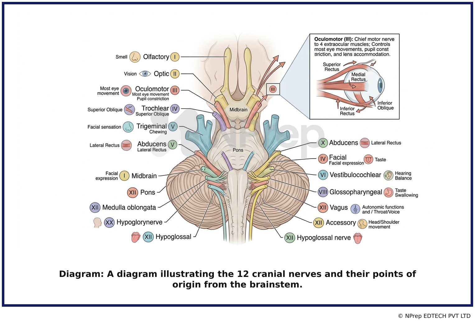
- Visual 1: Diagram: A diagram illustrating the 12 cranial nerves and their points of origin from the brainstem.
- Visual 2: Infographic: An infographic showing the six extraocular muscles of the eye, with labels indicating which cranial nerve (III, IV, or VI) innervates each muscle.
- Visual 3: Flowchart: A flowchart depicting the pathway of the Oculomotor nerve from its nucleus in the midbrain to the muscles it supplies in the orbit.
Clinical Relevance
- Nursing practice connection: This is primarily an exam-oriented knowledge point with limited direct bedside application, so retain Functions of Cranial Nerves as background academic context rather than a clinical decision trigger.
- A key nursing assessment is checking cranial nerve function. For CN III, this involves observing for ptosis (drooping eyelid), checking pupillary response to light, and assessing the six cardinal fields of gaze.
- Damage to the Oculomotor nerve (CN III palsy) can result in a characteristic 'down and out' position of the eye, a dilated pupil, and a drooping eyelid. This can be a sign of serious underlying conditions like a brain aneurysm, tumor, or increased intracranial pressure.
- What if the patient can move their eye laterally but not medially, up, or down? This specific deficit strongly suggests an isolated Oculomotor (CN III) nerve palsy, as the lateral rectus muscle (controlled by CN VI) is still functioning.
How to Approach the Question
- Identify the key function in the question stem: 'eye movement'.
- Recall the cranial nerves and their primary functions.
- Recognize that three nerves (III, IV, VI) control eye movement, but the question asks for the 'primary' one.
- Remember the mnemonic (LR6SO4)3: Lateral Rectus is CN VI, Superior Oblique is CN IV, and the rest (3) are CN III. Since CN III controls the most muscles, it is the primary nerve for eye movement.
- Evaluate the options based on this knowledge. Oculomotor (III) directly matches the function. The other options (Olfactory, Trigeminal, Vestibulocochlear) have distinct functions unrelated to eye movement.
Concept Tested & Keywords
- Concept Tested: Functions of Cranial Nerves
- Stem keywords: cranial nerve, eye movement
- Lead-in keywords: primarily responsible
Question ID
QZCGWHW_stHJDtBcHjLUqu
Practise the full KGMU 2023
Attempt every question from this paper in a timed mock, then review the full solution for each one.
More Nervous System Questions
- One function of parasympathetic nervous system is:
- What is the normal Intracranial pressure?
- Lumbar puncture for adults is done at:
- In healthy adults, the normal plantar flexion causes a:
- Which are the main functions of the hypoglossal nerve?
- Which is mainly responsible for fight (or) flight reactions?
- Nervous tissue is made of \ \ \ \ that receive and conduct impulses
- Which cortical area is responsible for the formulation of speech?
- Which reflex should check when death of the brain stem occurs, except?
- A patient is not able to sleep. On duty nurse what will suggest to do him?
More KGMU 2023 Questions
- The teacher scalded Aruna in front of her classmates during Project submission. Conclusion I: Aruna was not as popular as her other classmates. Concl…
- Aruna leaves and goes 8 meters in the North direction from a certain point. Then she turns right and travels for 12 meters. She again turns right and…
- Kiran is Sister of Raj. Raj is Son of Suresh and Vinita is Mother of Suresh then what is relation between Vinita and Kiran?
- A person who lives at the same time with another?
- Select the most appropriate meaning of the below idiom. A black sheep?
- The train has arrived...the station.
- A vendor sold 20 kg of potato at 18 Rs. per kg. He bought 2-liter milk at the rate of 46 Rs./liter, 2 kg sugar at the rate of 35 Rs./kg and 10 packet…
- Which one is the perfect square?
- A is the smallest. C is taller than B and D. E is neither the tallest nor the smallest. Find which one is the tallest?
- From an entrance, a person walks 28 meters left, then turns right and walks 13 meters. They turn right again and walk 12 meters, and then walk 16 met…