
NORCET-11 Prelims
Child Health Nursing (Pediatrics)
Medium
The given diagram illustrates the three main anatomical variants of neural tube defects Identify the correct sequential order of the types shown?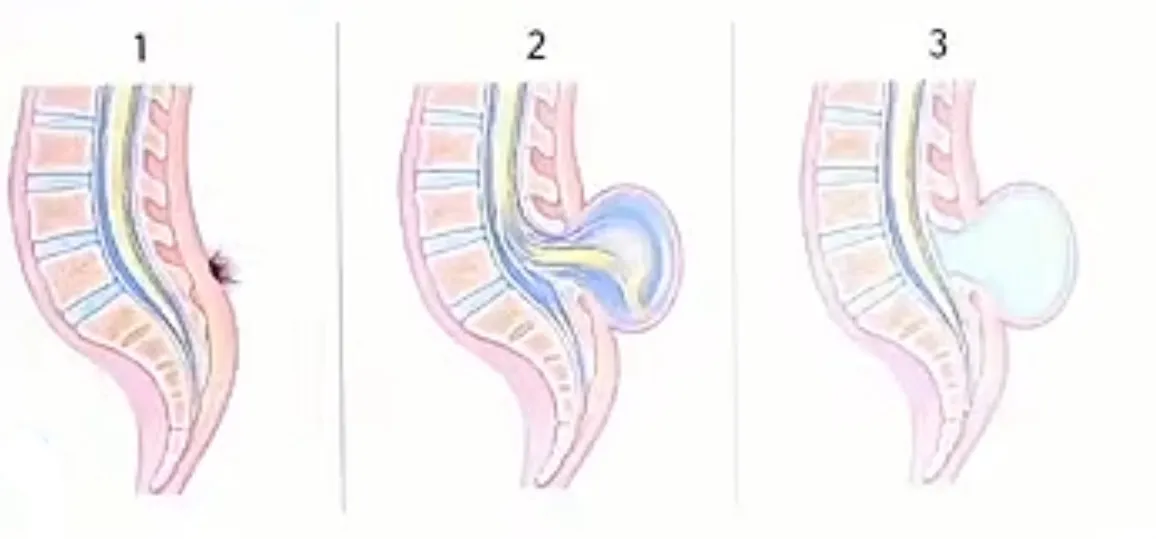
Appeared in: NORCET-11 Prelims
Explanation
- The first image shows spina bifida occulta: only vertebral arch defect, no sac, often a hairy patch.
- The second image shows myelomeningocele: sac contains CSF and meninges,with neural tissue.
- The third image shows meningocele: sac contains CSF, meninges, and no neural tissue.
- This order matches the increasing severity and anatomical involvement.
Why Other Options Were Wrong
- Option A: This sequence incorrectly places meningocele before mylomeningocele, which does not match the anatomical progression given show in image.
- Option C: This sequence starts with meningocele, which is not the mildest form. Spina bifida occulta should come first.
- Option D: This sequence starts with myelomeningocele, which is the most severe form, not the mildest.
Related Visual

Clinical Relevance
- Nursing practice connection: This is primarily an exam-oriented knowledge point with limited direct bedside application, so retain Identification and sequential classification of neural tube defect variants using anatomical images as background academic context rather than a clinical decision trigger.
- Nurses must recognize the type of neural tube defect to anticipate complications and plan care, especially for infection prevention and neurological assessment.
- Early identification helps guide surgical timing and parental counseling.
- What if? If the sac contains only CSF and meninges (no neural tissue), the child is less likely to have neurological deficits compared to myelomeningocele.
How to Approach the Question
- Carefully observe the anatomical features in each image panel.
- Recall the defining characteristics of each spina bifida type.
- Arrange the types from least to most severe based on neural involvement.
- Match the visual findings to the correct sequential order in the options.
- Eliminate options that do not follow the anatomical progression.
Concept Tested & Keywords
- Concept Tested: Identification and sequential classification of neural tube defect variants using anatomical images
- Stem keywords: diagram, neural tube defects, anatomical variants, sequential order
- Lead-in keywords: Identify, correct sequential order
- Clinical cues: Image shows progression from mild to severe neural tube defect
Question ID
QTJwy9WgRg0S7PTJTMJxzO
Reference Book
E6 Text Book Of Pediatric Nursing 3rd Panchali Pal — Part 2 (pp 239-476 of 713) p. 222-224
E6 Nelson Textbook of Pediatrics(2024) — Volume 2 p. 1368-1370
Practise the full NORCET-11 Prelims
Attempt every question from this paper in a timed mock, then review the full solution for each one.
More Central Nervous System Questions
- What is the most serious complication of meningitis in young children?
- A nurse is taking care of child who is having epilepsy. What nurse should do immediately?
- Which of the following nursing intervention can be used to prevent increased Intracranial pressure (ICP) in an unconscious child?
- Which of the following is NOT a complication of measles?
- Which of the following conditions can result in a hairy patch on the back of children?
- Which of the following symptoms suggest that the child has severe dehydration?
- Hypertonicity and scissoring leg movements are expected clinical findings in?
- Priority nursing action while caring for an infant with meningocele?
- What cranial nerve abnormality may be seen in children with chronic hydrocephalus?
- Neonates exposed to oxygen for a longer period may lead to?
More NORCET-11 Prelims Questions
- What is the capital of Arunachal Pradesh?
- A patient opens his eyes only when periorbital pressure is applied, makes incomprehensible sounds, and shows abnormal flexion to painful stimulus. Wh…
- Select the correctly spelt word from the options given below.
- Select the correctly spelt word from the options given below.
- Convert a patient's HbA1c value of 7% into estimated average glucose in mg/dL.
- A patient's recorded blood pressure is 150/90 mmHg. What is the calculated Mean Arterial Pressure?
- Identify the ECG pattern shown in the image below.
- At 36 weeks of pregnancy, a woman presents for an antenatal visit with suspected rupture of membranes. A Nitrazine paper test is conducted to differe…
- What is the official currency of Iran?
- Which of the following conditions is caused by a protozoan parasite?