
DSSSB 6 September 2024
Medical Surgical Nursing
Medium
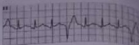
The ECG image given below represent
Appeared in: DSSSB 6 September 2024
Explanation
- The ECG shows a premature beat that interrupts the underlying normal sinus rhythm.
- The QRS complex of this premature beat is wide (greater than 0.12 seconds) and has a bizarre morphology, which is the hallmark of a ventricular origin.
- There is no P wave preceding the wide complex, confirming the impulse did not originate in the atria.
- A full compensatory pause follows the beat, which is characteristic of a PVC.
- These features collectively identify the arrhythmia as Premature Ventricular Contractions.
Why Other Options Were Wrong
- Option A: Premature atrial contractions (PACs) originate in the atria and are characterized by a premature P wave followed by a narrow QRS complex. The image shows a wide QRS complex with no preceding P wave.
- Option B: Atrial fibrillation is an irregularly irregular rhythm with no discernible P waves and a chaotic baseline. The ECG in the image shows a regular underlying rhythm with clear P waves for the normal beats.
- Option C: Atrial flutter is characterized by distinctive 'sawtooth' flutter waves in place of P waves. The baseline in the provided ECG is flat between the normal complexes, with no sawtooth pattern.
Related Visual

Clinical Relevance
- Nursing practice connection: This is primarily an exam-oriented knowledge point with limited direct bedside application, so retain ECG interpretation of ventricular arrhythmias as background academic context rather than a clinical decision trigger.
- While isolated PVCs are common and often benign in healthy individuals, frequent PVCs can be a sign of underlying heart disease, ischemia, or electrolyte imbalances (hypokalemia, hypomagnesemia).
- Nurses must assess the patient's clinical status, not just the monitor. Check for symptoms like palpitations, chest pain, or dizziness, as frequent PVCs can decrease cardiac output and blood pressure.
- It is critical to report the frequency and pattern of PVCs. Runs of three or more consecutive PVCs constitute ventricular tachycardia (VT), a life-threatening arrhythmia requiring immediate intervention.
How to Approach the Question
- First, analyze the overall rhythm. Look at the R-R intervals of the normal-looking beats to determine if the underlying rhythm is regular or irregular.
- Next, identify the components of the normal beats: find the P wave, measure the PR interval, and measure the QRS duration (narrow or wide). This establishes the baseline rhythm (in this case, normal sinus rhythm).
- Scan the strip for any beats that look different or occur early. In this image, one beat clearly stands out.
- Analyze the abnormal beat: Is it early? Is there a P wave before it? Is the QRS complex narrow or wide and bizarre?
- Synthesize the findings: An early beat with no P wave and a wide, bizarre QRS complex is the classic presentation of a Premature Ventricular Contraction (PVC).
- Compare these findings to the definitions of the other options (PAC, A-fib, A-flutter) to confirm they do not match.
Concept Tested & Keywords
- Concept Tested: ECG interpretation of ventricular arrhythmias
- Stem keywords: ECG image
- Lead-in keywords: represent
- Clinical cues: Presence of an ectopic beat on an ECG strip
- Negative lead-in flag: false
Question ID
Qi100SwYJaT0ebLg9MBdiX
Reference Book
E6 Medicine Harrison 22e Part 1 p. 2001-2003
E6 Pharmacology Nursing Lilley 11e Part 2 p. 86-88
E6 Nelson Textbook of Pediatrics(2024) — Volume 2 p. 633-635
Practise the full DSSSB 6 September 2024
Attempt every question from this paper in a timed mock, then review the full solution for each one.
More ECG Questions
More DSSSB 6 September 2024 Questions
- Which of the following is NOT a primitive defence mechanism?
- After taking lithium drug for one month, a patient checked their serum lithium level and found it to be 1.0 mEq/L. The nurse checked the report and n…
- Select the option that correctly describes the condition being shown in the following image.
- Turbid, cloudy CSF is commonly associated with:
- According to the Mental Health Act, 1987, who from the following make(s) the application for admission under special circumstances?
- Which of the following types of schizophrenia is commonly characterised by excitement and stupor?
- What is the action of the Sitagliptin?
- Which gas is most commonly used for daily work in the wards and operation theatres?
- Sickle cell RBCs are also known as:
- Glimepiride is an example of a \ \ \ \ \ \ .