
KGMU 2023
Applied Physiology
Easy
In which part of the human eye is the image of an object formed?
Appeared in: KGMU 2023
Explanation
- The retina is the innermost, light-sensitive layer of the eyeball, analogous to the film or sensor in a camera.
- It contains specialized photoreceptor cells, known as rods and cones, which are essential for vision.
- Light entering the eye is focused by the cornea and lens to form a sharp, inverted image directly on the retina.
- These photoreceptor cells then convert the light energy into electrical signals, which are transmitted to the brain via the optic nerve for interpretation.
Why Other Options Were Wrong
- Option B: The cornea is the transparent front part of the eye that provides the most significant refraction (bending) of light, but it is not where the final image is formed.
- Option C: The iris is the colored muscular diaphragm that controls the size of the pupil, thereby regulating the amount of light that enters the eye. It does not participate in image formation.
- Option D: The sclera is the tough, white, fibrous outer layer that provides structural support and protection for the eyeball. It is opaque and not involved in the visual pathway.
Related Visual
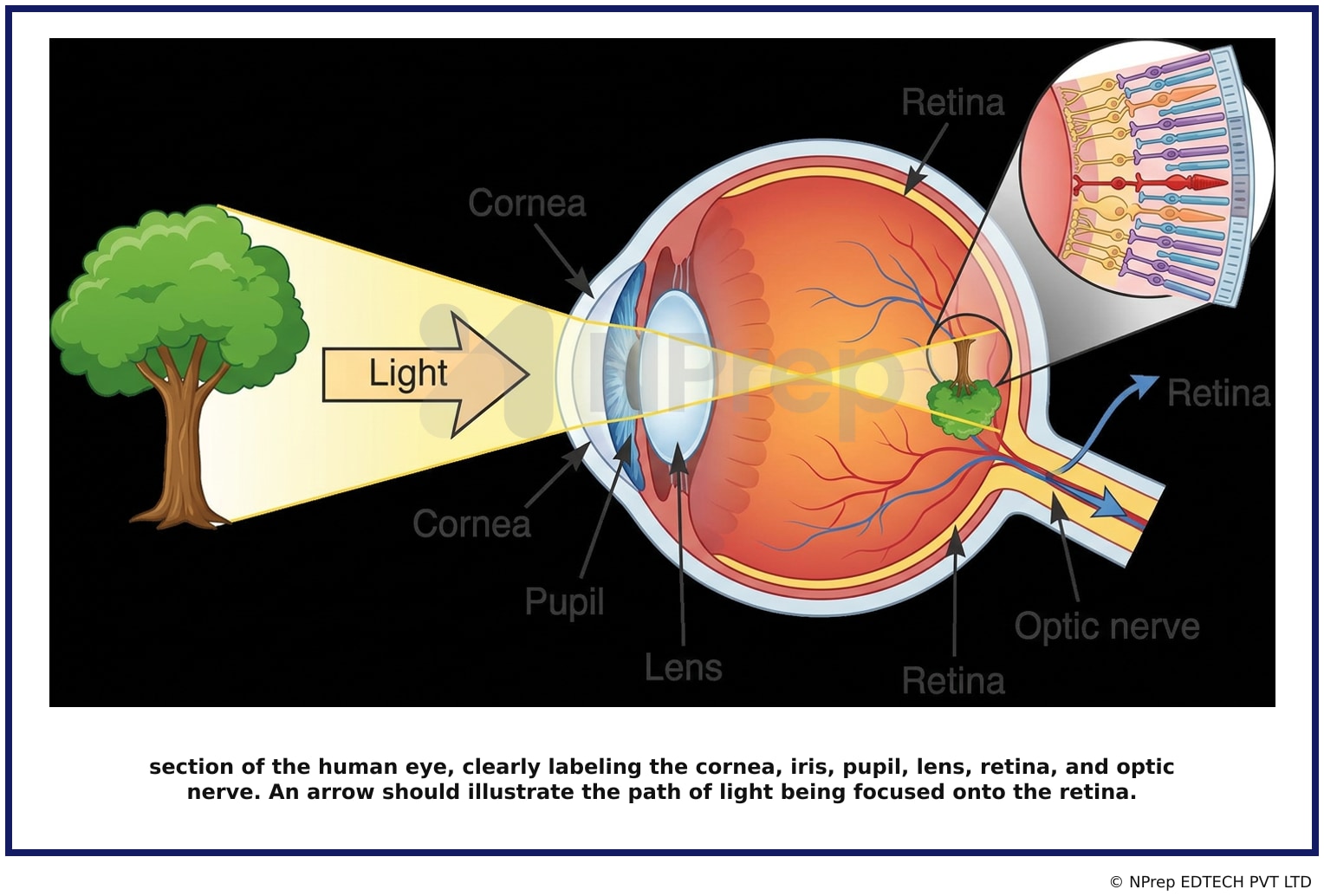
- Visual 1: Diagram - A cross-section of the human eye, clearly labeling the cornea, iris, pupil, lens, retina, and optic nerve. An arrow should illustrate the path of light being focused onto the retina.
Clinical Relevance
- Nursing practice connection: This is primarily an exam-oriented knowledge point with limited direct bedside application, so retain Anatomy and function of the human eye, specifically the site of image formation as background academic context rather than a clinical decision trigger.
- Understanding that the image forms on the retina is fundamental to diagnosing and explaining refractive errors. In myopia (nearsightedness), the image is focused in front of the retina, while in hyperopia (farsightedness), it is focused behind it.
- Many serious eye diseases, such as diabetic retinopathy, macular degeneration, and retinal detachment, directly affect the retina's ability to receive an image, leading to vision loss.
- What if? If a patient experiences a retinal detachment, the retina physically separates from the underlying choroid layer. This prevents a clear image from being formed, causing symptoms like a 'curtain' over the vision, sudden floaters, and flashes of light. It is a medical emergency requiring immediate surgery to prevent permanent blindness.
How to Approach the Question
- First, identify the key concept in the question: 'where the image is formed' in the eye.
- Recall the basic function of the eye as an optical system similar to a camera, which requires a lens to focus light and a sensitive surface to capture the image.
- Mentally trace the path of light: It enters through the transparent cornea, passes through the pupil (controlled by the iris) and the lens, and is finally focused.
- Evaluate each option based on its primary function: The cornea bends light, the iris controls light quantity, and the sclera protects the eye.
- Conclude that the retina is the only structure listed that is a light-sensitive layer designed to detect the focused image and convert it into neural signals.
Concept Tested & Keywords
- Concept Tested: Anatomy and function of the human eye, specifically the site of image formation.
- Stem keywords: human eye, image, formed
- Lead-in keywords: In which part
- Negative lead-in flag: false
Question ID
QNdsGEJYGaKgV_4SkK7I2r
Practise the full KGMU 2023
Attempt every question from this paper in a timed mock, then review the full solution for each one.
More Sensory organs Questions
- Which of the following helps prevent water loss through skin?
- Which of the following response is anticipated in a mother during suckling the baby?
- Anosmia is?
- Which is transmission of sound vibrations to the internal ear?
- What is the normal intraocular pressure in an adult?
- The size of which part of the eye contributes to controlling the amount of light entering the eye?
- The main function of sweda is:
- It is a type of refractive error in which light rays are focused in front of the retina?
- Which of the following photoreceptors helps in "bright light vision"?
- The Rods and Cones of the eye retinal layer are modified?
More KGMU 2023 Questions
- The teacher scalded Aruna in front of her classmates during Project submission. Conclusion I: Aruna was not as popular as her other classmates. Concl…
- Aruna leaves and goes 8 meters in the North direction from a certain point. Then she turns right and travels for 12 meters. She again turns right and…
- Kiran is Sister of Raj. Raj is Son of Suresh and Vinita is Mother of Suresh then what is relation between Vinita and Kiran?
- A person who lives at the same time with another?
- Select the most appropriate meaning of the below idiom. A black sheep?
- The train has arrived...the station.
- A vendor sold 20 kg of potato at 18 Rs. per kg. He bought 2-liter milk at the rate of 46 Rs./liter, 2 kg sugar at the rate of 35 Rs./kg and 10 packet…
- Which one is the perfect square?
- A is the smallest. C is taller than B and D. E is neither the tallest nor the smallest. Find which one is the tallest?
- From an entrance, a person walks 28 meters left, then turns right and walks 13 meters. They turn right again and walk 12 meters, and then walk 16 met…