
AIIMS Delhi NO - 2018
Child Health Nursing (Pediatrics)
Easy
Identify the disorder in the image below?
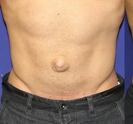
Appeared in: AIIMS Delhi NO - 2018
Explanation
- The image clearly shows a protrusion at the umbilicus (belly button), which is the defining characteristic of an umbilical hernia.
- This type of hernia results from the incomplete closure of the umbilical ring after birth or a weakening of the abdominal wall in adults.
- The hernia appears as a soft swelling covered by skin that may become more prominent when intra-abdominal pressure increases, such as during crying, coughing, or straining.
Why Other Options Were Wrong
- Option A: An inguinal hernia presents as a bulge in the groin area, not at the navel. The image does not show a swelling in the groin.
- Option B: A femoral hernia appears as a lump in the upper thigh or groin, just below the inguinal ligament. The bulge in the image is located centrally on the abdomen at the navel.
- Option D: A diaphragmatic hernia is an internal condition where abdominal organs protrude into the chest cavity through a defect in the diaphragm. It does not produce a visible external bulge on the abdomen and is typically diagnosed through imaging and respiratory symptoms.
Related Visual

Clinical Relevance
- Nursing practice connection: This is primarily an exam-oriented knowledge point with limited direct bedside application, so retain Visual identification of abdominal hernias as background academic context rather than a clinical decision trigger.
- Nurses must be able to visually identify different types of hernias to perform an accurate initial assessment and report findings correctly. The location of the bulge is the primary clue.
- Patient education is crucial. For umbilical hernias, nurses should advise patients to avoid heavy lifting and straining, which can increase abdominal pressure and worsen the hernia.
- A key nursing responsibility is to assess for signs of strangulation (incarceration), where the blood supply to the herniated tissue is cut off. Symptoms include sudden, severe pain, redness, nausea, and vomiting. This is a medical emergency.
How to Approach the Question
- First, carefully observe the provided image to identify the key clinical sign. Note the precise location of the abnormality on the patient's body.
- In this case, the key feature is a prominent bulge located directly at the umbilicus (belly button).
- Next, review the provided options, which list different types of hernias.
- Systematically evaluate each option based on your anatomical knowledge. An inguinal hernia is in the groin, a femoral hernia is in the upper thigh, and a diaphragmatic hernia is internal.
- Match your observation from the image to the correct definition. The bulge at the umbilicus directly corresponds to the definition of an umbilical hernia.
- Select the option that accurately describes the visual evidence.
Concept Tested & Keywords
- Concept Tested: Visual identification of abdominal hernias
- Stem keywords: disorder, image
- Lead-in keywords: Identify
- Clinical cues: Visible bulge at the umbilicus (navel)
Question ID
QH08rAaTm3hq7hhL_iaeaE
Reference Book
E6 Nelson Textbook of Pediatrics(2024) — Volume 1 p. 1142-1144
E6 Text Book Of Pediatric Nursing 3rd Panchali Pal — Part 2 (pp 239-476 of 713) p. 169-171
Practise the full AIIMS Delhi NO - 2018
Attempt every question from this paper in a timed mock, then review the full solution for each one.
More Normal Growth and its Disorders Questions
- What is the scoring system to estimate the gestational age of a newborn by maturity rating?
- The infant's weight becomes triple from birth weight in:
- Number of teeth in a 3-year old child is:
- When does the infant's weight get double in relation to birth weight?
- A kind of play which can be observed in infants:-
- The term 'Cephalocaudal' means:-
- At what time, an average term baby doubles the birth weight? (Choose the appropriate option from below)
- During physical assessment of children, asymmetry of the skull indicates?
- Which of the following is NOT true regarding the birth weight of a newborn?
- In the stages of growth and development, infancy extends from?
More AIIMS Delhi NO - 2018 Questions
- Another tentative diagnosis made for the client is Wernicke's Encephalopathy. If the client has this, the nurse anticipates that the first physician'…
- A 35-year-old man was in an automobile accident with an admitting diagnosis of a head injury. He has a Glasgow Coma Scale score of 4. Upon MRI and CT…
- A 28-year-old male client is admitted to the hospital for a suspected brain tumor. While assessing this client, the nurse would keep in mind that the…
- Following admission to the hospital, a diagnosis of increasing intracranial pressure was made. The nursing intervention appropriate in the care of th…
- The nurse has orders to administer phenytoin (Dilantin) 100 mg IV as an anticoagulant. The priority action while administering this drug is to:
- The client is to take lithium regularly after she is discharged from the hospital. The nursing care plan includes discharge planning. The most import…
- A patient is taking a proper diet as prescribed for bulimia nervosa, but the patient's weight is not gaining. Which nursing action is appropriate?
- Identify the size of the cannula shown here?& x20;
- Identify the following image? & x20;
- Identify the instrument below?& x20;