
OSSSC Nursing Officer-2021
Obstetrics and Midwifery Nursing
Easy
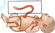
Cut-section of Structure shown in the photograph contains
Appeared in: OSSSC Nursing Officer-2021
Explanation
- The image displays a newborn with an umbilical cord, which is the structure in question.
- A standard, full-term umbilical cord contains three blood vessels.
- Specifically, there are two umbilical arteries and one umbilical vein.
- The two arteries carry deoxygenated blood away from the fetus.
- The single vein carries oxygenated, nutrient-rich blood to the fetus.
- This 2-artery, 1-vein composition is the correct anatomical configuration.
Why Other Options Were Wrong
- Option A: This option incorrectly states there is one artery and two veins. While two umbilical veins exist very early in embryonic development, the right vein typically degenerates, leaving only one.
- Option B: This option suggests two arteries and two veins. A term umbilical cord does not normally contain four vessels.
- Option D: This option describes one artery and one vein. This is known as a Single Umbilical Artery (SUA) and is the most common malformation of the cord, but it is not the normal anatomy.
Related Visual

Clinical Relevance
- Nursing practice connection: This is primarily an exam-oriented knowledge point with limited direct bedside application, so retain Anatomy of the Umbilical Cord as background academic context rather than a clinical decision trigger.
- A nurse assisting with delivery must be able to identify a normal umbilical cord and note any abnormalities, such as a Single Umbilical Artery (SUA), as it may indicate a higher risk for other congenital defects.
- Post-delivery, the nurse inspects the clamped cord for three vessels. The finding of only two vessels (SUA) must be documented and communicated to the pediatric team for further evaluation of the newborn.
- What if? If an ultrasound at 20 weeks revealed a single umbilical artery, the obstetrician would recommend a detailed fetal anatomical survey and possibly a fetal echocardiogram to screen for associated cardiac and renal anomalies.
How to Approach the Question
- First, identify the structure shown in the photograph. The image clearly shows a newborn baby with its umbilical cord.
- The question asks for the contents of a 'cut-section' of this structure, which means you need to know the internal anatomy of the umbilical cord.
- Recall the normal anatomical composition of a human umbilical cord. It serves as the connection between the fetus and the placenta.
- Remember the mnemonic 'AVA' which stands for Artery-Vein-Artery, indicating two arteries and one vein.
- Evaluate the given options based on this anatomical fact.
- Select the option that matches the normal composition: 2 arteries and 1 vein.
Concept Tested & Keywords
- Concept Tested: Anatomy of the Umbilical Cord
- Stem keywords: Cut-section, Structure, photograph
- Lead-in keywords: contains
- Clinical cues: Image of a newborn with an umbilical cord
Question ID
q1rpbnU1W6nGoC8fckqrJ
Reference Book
E6 Obstetrics Williams p. 71-95
E6 Histology Inderbir 11e p. 5-7
E6 Nelson Textbook of Pediatrics(2024) — Volume 1 p. 1141-1143
Practise the full OSSSC Nursing Officer-2021
Attempt every question from this paper in a timed mock, then review the full solution for each one.
More Placenta and Fetal Membranes Questions
- The following are the characteristics of fetal umbilical cord; EXCEPT:
- Formation of the placenta and full functioning of the placenta develops at?
- The umbilical cord at term contains?
- The approximate average weight of the placenta at birth is?
- Substance that crosses the placental barrier is?
- During intrauterine life, the main source of nutrition to the fetus is?
- How much amount of blood transfers from placenta to the baby after delivery when the cord clamping is delayed till the cessation of cord pulsations?
- Which is the only immunoglobulin transported through placenta to fetus?
- In which condition, the umbilical cord is attached to the margins of the placenta?
- Which antibody crosses the placenta?
More OSSSC Nursing Officer-2021 Questions
- A man completed a journey in 10 hours. He travelled first half of the total distance at the rate of 21 km/hr and second half at the rate of 24 km/hr.…
- A person crosses a 600 metre long street in 5 minutes. What is his speed in kilometer per hour?
- What is the cube of -1 1/3?
- What is the ratio between 0.02 and 0.002?
- After 4 years the principal amount of Rs. 12000 grows into Rs. 14,400. Find out the rate of simple interest?
- What is the decimal of 9% ?
- If the marked price of an article is Rs. 400/- and its sale price is Rs. 340/-, Find out the percentage of discount.
- If three-fifth (3/5) of 60% of a number is 36, then what is the number?
- After 15 years, a man will be just four times as old as he was 15 years ago. What is his present age?