
MP CHO -2019 (Shift-1)
Applied Anatomy
Easy
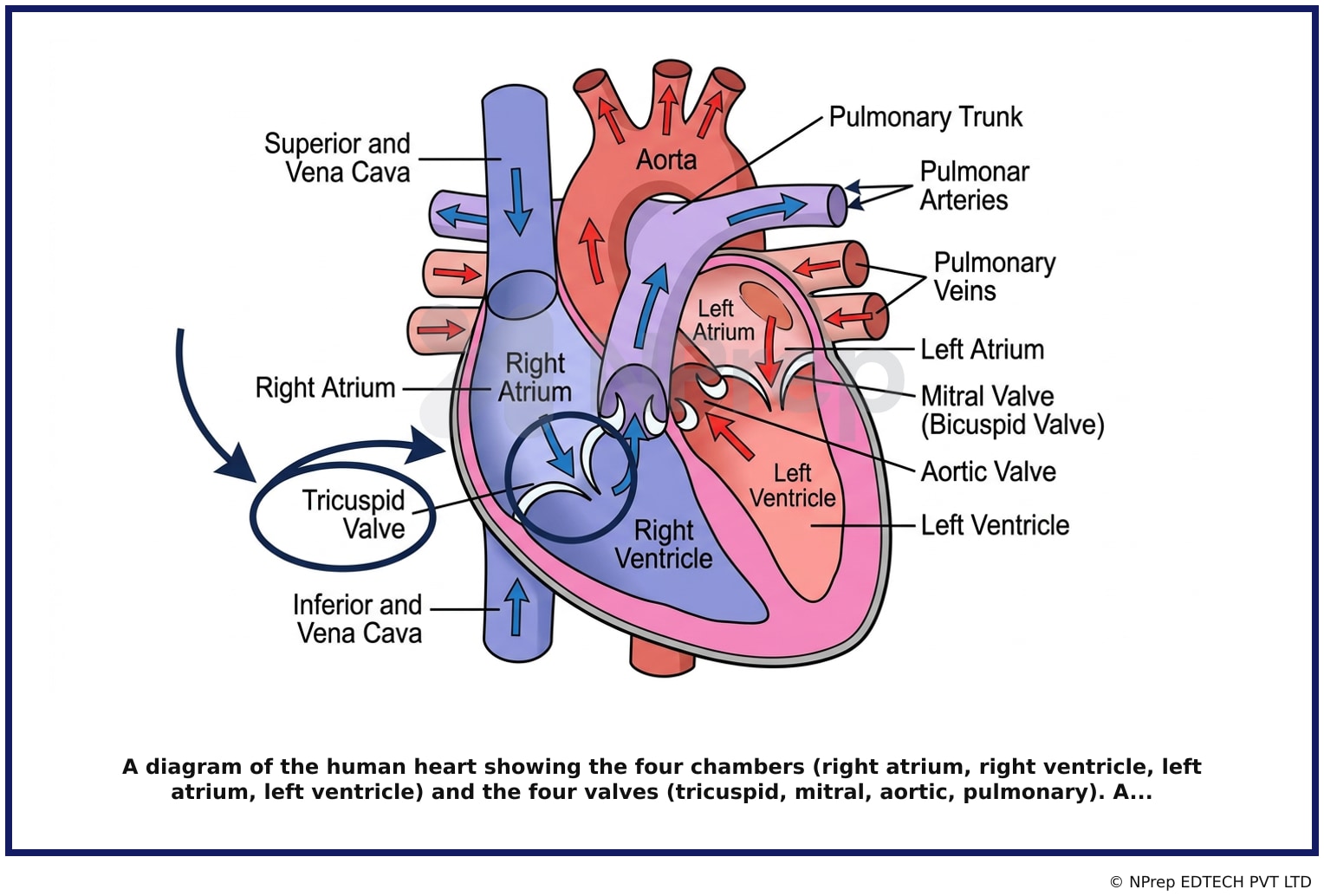
The opening of the right atrium into the right ventricle occurs through which valve?
Appeared in: MP CHO -2019 (Shift-1)
Explanation
- The tricuspid valve is situated between the right atrium and the right ventricle.
- It is an atrioventricular (AV) valve, named for its three leaflets (cusps).
- Its primary role is to open during atrial contraction to allow deoxygenated blood to fill the right ventricle and to close securely during ventricular contraction to prevent blood from flowing back into the right atrium.
Why Other Options Were Wrong
- Option A: The mitral valve is located on the left side of the heart, between the left atrium and the left ventricle. It controls the flow of oxygenated blood.
- Option C: The bicuspid valve is another name for the mitral valve. It is located between the left atrium and the left ventricle, not the right.
- Option D: The aortic semilunar valve is located at the exit of the left ventricle, opening into the aorta. It is not situated between an atrium and a ventricle.
Related Visual
Clinical Relevance
- Nursing practice connection: This is primarily an exam-oriented knowledge point with limited direct bedside application, so retain Anatomy and function of the heart valves, specifically the location of the tricuspid valve as background academic context rather than a clinical decision trigger.
- Auscultating heart sounds is a fundamental nursing assessment. The sound of the tricuspid valve closing (part of the S1 'lub' sound) is best heard at the left lower sternal border, in the 4th or 5th intercostal space.
- Conditions like tricuspid stenosis (narrowing) or regurgitation (leaking) can impair heart function, leading to symptoms like fatigue, swelling in the legs (edema), and abdominal swelling (ascites).
- What if? If a patient has tricuspid regurgitation, a systolic murmur would be heard because the valve fails to close properly, allowing blood to leak back into the right atrium when the right ventricle contracts.
How to Approach the Question
- First, identify the key anatomical structures mentioned in the question: the 'right atrium' and the 'right ventricle'.
- The question asks to identify the 'valve' that connects these two specific chambers.
- Recall the basic anatomy of the heart and the sequence of blood flow. Deoxygenated blood enters the right atrium, passes through a valve into the right ventricle, is pumped to the lungs, returns as oxygenated blood to the left atrium, passes through another valve to the left ventricle, and is then pumped to the body.
- Focus on the right side of the heart. The valve between the right atrium and right ventricle is the tricuspid valve.
- Evaluate the given options: Mitral/Bicuspid and Aortic valves are on the left side of the heart or exiting it. The tricuspid valve is the only option that correctly fits the location described.
- Select the option that names the tricuspid valve.
Concept Tested & Keywords
- Concept Tested: Anatomy and function of the heart valves, specifically the location of the tricuspid valve.
- Stem keywords: right atrium, right ventricle, opening, valve
- Lead-in keywords: which valve
Question ID
Q6rxzc732996T0UqEIK0hZ
Reference Book
E6 Anatomy Applied Ashalatha 4e pp. 165-167, 163-165, 164-166
Practise the full MP CHO -2019 (Shift-1)
Attempt every question from this paper in a timed mock, then review the full solution for each one.
More Circulatory & Lymphatic System Questions
- The 'pacemaker' of the heart is:-
- Most common venous access site in upper limbs?
- The apex of the heart is formed mostly by the?
- The main arterial stem of the upper limb is?
- Which artery supplies oxygenated blood to the stomach?
- Distal blood vessel of leg is?
- Pacemaker of heart is?
- JVP point is?
- How many chambers in the human heart?
- The pacemaker of the heart is?
More MP CHO -2019 (Shift-1) Questions
- Who among the following is the Governor of Madhya Pradesh?
- P. V. Sindhu is associated with which sport?
- Who is the Union Health Minister?
- When is World Health Day celebrated?
- Which of the following is not correct?
- Which of the following statements is correct?
- Which is the largest state in India?
- What is the largest internal organ in the human body?
- What is Rafale?
- Where is Darbar Sahib Kartarpur Gurdwara located?