
NORCET-7 Mains-2024
Obstetrics & Gynaecology
Easy
In the following USG scan snowstorm appearance is seen which is possibly seen in? 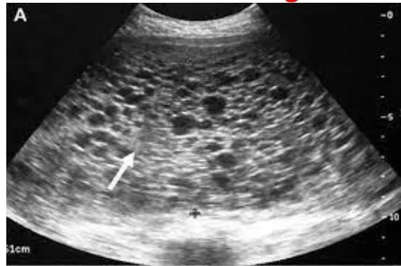
Appeared in: NORCET-7 Mains-2024
Explanation
- The ultrasound image displays a classic "snowstorm" pattern within the uterus, which is a pathognomonic (a characteristic sign) for a Hydatidiform Mole.
- A hydatidiform mole is a form of gestational trophoblastic disease where there is abnormal proliferation of the chorionic villi of the placenta.
- The "snowstorm" appearance is created by these numerous swollen, fluid-filled (hydropic) villi, which appear as multiple small cysts on the ultrasound.
- The absence of a fetus in the image is characteristic of a complete mole, which results from the fertilization of an empty egg.
Why Other Options Were Wrong
- Option A: An ectopic pregnancy involves implantation outside the uterine cavity. An ultrasound would typically show an empty uterus and a gestational sac or complex mass in an adnexal location (e.g., fallopian tube).
- Option C: Endometriosis involves endometrial tissue growing outside the uterus. On ultrasound, it may appear as ovarian cysts (endometriomas) with a uniform 'ground-glass' echo pattern, not a multicystic mass inside the uterus.
- Option D: Disseminated Intravascular Coagulation (DIC) is a systemic disorder of blood clotting and is not a structural abnormality diagnosed by ultrasound. It can be a severe complication of a molar pregnancy but is not the primary diagnosis based on this image.
Related Visual

- Visual 1: Diagram: Pathogenesis of a complete mole vs. a partial mole, showing the chromosomal origin (androgenesis in a complete mole).
- Visual 2: Chart: Typical pattern of serum beta-hCG level decline after successful evacuation of a molar pregnancy versus the pattern seen in persistent gestational trophoblastic neoplasia (GTN).
Clinical Relevance
- Nursing practice connection: This is primarily an exam-oriented knowledge point with limited direct bedside application, so retain Ultrasound diagnosis of Hydatidiform Mole as background academic context rather than a clinical decision trigger.
- Nurses must monitor patients with molar pregnancy for signs of serious complications, including hemorrhage, hyperthyroidism (due to the thyrotropin-like effects of hCG), and severe preeclampsia, which can occur early in gestation.
- Patient education is a critical nursing role, focusing on the necessity of long-term follow-up with serial beta-hCG monitoring to screen for malignant transformation into choriocarcinoma.
- It is vital to counsel the patient on the importance of using reliable contraception during the entire follow-up period, as a new pregnancy would produce hCG and interfere with monitoring for malignancy.
How to Approach the Question
- First, identify the type of image provided, which is a pelvic ultrasound (USG).
- Systematically analyze the image for key features. Observe the contents of the uterus, noting the heterogeneous texture and the presence of multiple small, black (anechoic) circles.
- Recognize this distinctive pattern as the classic "snowstorm" appearance.
- Recall or deduce which obstetrical condition is associated with this pathognomonic sign.
- Evaluate the given options. Hydatidiform mole is the direct match for the "snowstorm" sign.
- Rule out the other options by considering their typical, and different, sonographic presentations.
Concept Tested & Keywords
- Concept Tested: Ultrasound diagnosis of Hydatidiform Mole
- Stem keywords: USG scan, snowstorm appearance
- Lead-in keywords: possibly seen in
- Clinical cues: Image shows 'snowstorm' appearance on ultrasound, a pathognomonic sign.
Question ID
QpJlrX4x2MotOH2llRX8Do
Practise the full NORCET-7 Mains-2024
Attempt every question from this paper in a timed mock, then review the full solution for each one.
More Hemorrhage in Early Pregnancy Questions
- Nurse's main priority assessment while caring for the client having spontaneous abortion:
- Most common site of ectopic pregnancy?
- In ectopic pregnancy deciduas are shed as?
- The most common site of ectopic pregnancy is?
- Chief site of ectopic pregnancy is?
- When a fertilized ovum is implanted outside the uterine cavity, it is called?
- Which of the following is the method of termination of pregnancy in the second trimester (13 to 20 weeks)?
- A woman with 36 weeks of gestation informs the nurse there is a sudden gush of fluid from the vagina. This indicates which of the clinical conditions?
- What is the type of abortion wherein the fetus is dead but retained inside the uterus for a variable period?
- Which of the following is NOT a characteristic of a threatened abortion?
More NORCET-7 Mains-2024 Questions
- What is the correct statement regarding pre-oxygenation in children before intubation?
- A pregnant woman is ready for caesarean section and transferred to labor room. Which is the best position for the woman during caesarean section?
- A woman comes in labor room with 3 cm cervical dilation, 80% effacement and energetic and excited. The women in which phase of labor?
- The ambient temperature of the labor room should maintain?
- The recommended dose of iron and folic acid for adolescent girls under Weekly Iron & Folic Acid Supplementation (WIFS) program?
- A 14-week Rh-negative pregnant woman presents with mild vaginal bleeding and abdominal pain. Ultrasound shows a viable intrauterine pregnancy. Her hu…
- A new newborn care unit is inaugurated in the hospital. The child health nurse known about iKMC. What mean I in iKMC?
- A nurse teaching to a woman regarding breast feeding. Which statement is incorrect about breast feeding?
- Swenson and Duhamel procedure used to correction of?
- Cohort study also known as?