
INI-CET EXAM -2025
Applied Anatomy
Medium
Identify the nerve block demonstrated in the given intraoral image.

Appeared in: INI-CET EXAM -2025
Explanation
- The image clearly shows the needle tip being placed in the hard palate, medial to the maxillary posterior teeth.
- This specific anatomical location is the site of the greater palatine foramen.
- The greater palatine nerve emerges from this foramen to supply sensation to the posterior hard palate.
- Therefore, injecting anesthetic at this site constitutes a greater palatine nerve block, which anesthetizes the palatal mucosa and tissues from the third molar to the first premolar area.
Why Other Options Were Wrong
- Option A: The inferior alveolar nerve block is administered in the mandible (lower jaw) to anesthetize the lower teeth, lip, and chin. The image clearly shows an injection in the maxilla (upper jaw).
- Option B: The anterior ethmoidal nerve block anesthetizes parts of the nasal cavity. The injection is given intranasally or extraorally, not on the hard palate as shown.
- Option C: A maxillary nerve block is a more extensive, regional block that anesthetizes an entire half of the maxilla. It is administered at a higher anatomical site, such as the pterygopalatine fossa, not as a simple injection on the palate.
Related Visual
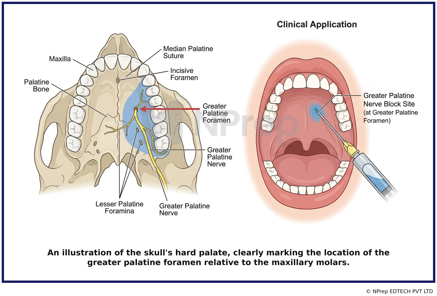
- Visual 1: Diagram - An illustration of the skull's hard palate, clearly marking the location of the greater palatine foramen relative to the maxillary molars.
- Visual 2: Flowchart - A chart showing the branches of the maxillary nerve (V2) and the areas they innervate, highlighting the greater palatine nerve's distribution on the palate.
- Visual 3: Infographic - A comparative infographic showing the needle placement and area of anesthesia for the four nerve blocks listed as options.
Clinical Relevance
- Nursing practice connection: This is primarily an exam-oriented knowledge point with limited direct bedside application, so retain Identification of local anesthetic nerve block techniques in dentistry as background academic context rather than a clinical decision trigger.
- The greater palatine nerve block is a fundamental technique for achieving pain control during dental procedures involving the palatal tissues of the maxillary posterior teeth.
- It is commonly used for extractions, periodontal surgery, or when a rubber dam clamp needs to be placed on a posterior tooth, which might impinge on the palatal gingiva.
- Nurses and dental assistants must be able to recognize the procedure to prepare the correct armamentarium and anticipate the area of anesthesia for patient monitoring.
How to Approach the Question
- First, analyze the image to identify the anatomical context. Observe that the injection is in the oral cavity, specifically the upper jaw (maxilla) and on the roof of the mouth (palate).
- Next, pinpoint the specific landmark. The needle is directed towards the posterior part of the hard palate, near the molar teeth.
- Recall the common dental nerve blocks and their target locations. The greater palatine foramen is located in this exact area.
- Evaluate the given options. The 'Inferior alveolar' block is for the lower jaw. The 'Anterior ethmoidal' is for the nose. The 'Maxillary' block is a high-level block with a different approach.
- Conclude that the only option matching the visual evidence of an injection at the greater palatine foramen is the 'Greater palatine nerve block'.
Concept Tested & Keywords
- Concept Tested: Identification of local anesthetic nerve block techniques in dentistry.
- Stem keywords: nerve block, intraoral image, identify
- Lead-in keywords: Identify
- Clinical cues: The image shows a needle directed at the hard palate of the upper jaw, which is a key visual cue.
Question ID
QufFabvRwDmU2ZeJ5S2jtE
Practise the full INI-CET EXAM -2025
Attempt every question from this paper in a timed mock, then review the full solution for each one.
More Nervous System Questions
- Which one of the following is the third cranial nerve?
- Identify the procedure done by the health care professional in the image below:
- Connection between right and left hemisphere is known as?
- A young adult who was hit by a car was fortunate because the level of his injury did not interrupt his respiratory function. The cord segment involve…
- Auditory area located in which lobe of the brain?
- Following a head injury, a person comes into the emergency department with personality changes and loss of voluntary control. Which lobe of the brain…
- Pressure by the upper end of a crutch may cause injury to the?
- Carpal tunnel syndrome is caused by the compression of?
- Which of the following is the largest cranial nerve?
- How many cervical nerves are there in the spinal cord?
More INI-CET EXAM -2025 Questions
- The highlighted structure in the provided image articulates with which bone?& x20;
- Through which of the following foramina does the nerve supplying the muscles of mastication pass?& x20;
- In the given image, which region is correctly paired with its corresponding brain structure? • Middle cerebellar peduncle • Fasciculus cuneatus • Sup…
- Which of the following muscles is NOT typically associated with dysphagia or dysarthria?
- A muscle of mastication involved in opening the mouth exhibits the following characteristics. Identify the incorrect statement about it:
- In the illustrated image, which of the following structures is NOT associated with the area marked by the arrow?& x20;
- Match the following embryonic aortic arches with their respective adult derivatives: A. 1st Arch 1. Maxillary artery& x20; B. 2nd Arch 2. Common caro…
- Which of the following histology image is not an endocrine structure?& x20;
- Marked structure separates which of the following:
- Which of the following bones of the skull gives passage to a nerve arising from the brain & x20;