
AIIMS Jodhpur SNO-2023
Applied Anatomy
Easy
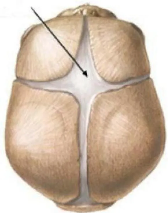 Identify the fontanel indicated by the arrow.
Appeared in: AIIMS Jodhpur SNO-2023
Explanation
- The image displays a superior view of an infant's skull, with the arrow pointing to the anterior fontanel.
- This fontanel is characteristically large and diamond-shaped, which is a key visual identifier.
- It is located at the junction of the two frontal bones and the two parietal bones, where the coronal and sagittal sutures meet.
- Clinically, it is the last major fontanel to close, typically between 12 and 18 months of age.
Why Other Options Were Wrong
- Option B: The posterior fontanel is located at the back of the skull, is triangular in shape, and is much smaller than the structure shown.
- Option C: The temporal (or sphenoidal) fontanel is located on the side of the skull, in the temple region, not on the top.
- Option D: The occipital fontanel is another name for the posterior fontanel. The structure shown is the anterior fontanel due to its diamond shape and anterior location.
Related Visual

Clinical Relevance
- Nursing practice connection: This is primarily an exam-oriented knowledge point with limited direct bedside application, so retain Anatomical identification of infant skull fontanels as background academic context rather than a clinical decision trigger.
- Assessing fontanels is a fundamental part of the neonatal and infant physical examination. It provides vital clues about hydration status and neurological condition.
- A sunken or depressed fontanel in an infant is a classic sign of dehydration and requires prompt fluid replacement.
- A tense or bulging fontanel when the infant is calm is a red flag for increased intracranial pressure (ICP), which can be caused by meningitis, hydrocephalus, or intracranial hemorrhage, and constitutes a medical emergency.
How to Approach the Question
- First, carefully examine the image provided. Note the view of the skull (superior/top-down) and the specific location the arrow is pointing to.
- Identify the key anatomical features of the indicated structure. In this case, observe its large size and distinct diamond shape.
- Recall the names and characteristics of the different fontanels in an infant's skull: anterior, posterior, sphenoidal (temporal), and mastoid.
- Compare the features seen in the image with your knowledge. The diamond shape and anterior location are characteristic of the anterior fontanel.
- Evaluate the given options. 'Anterior fontanel' directly matches your identification. 'Posterior' and 'Occipital' refer to the same triangular fontanel at the back. 'Temporal' refers to the fontanel on the side.
- Select the option that correctly identifies the structure based on the visual evidence.
Concept Tested & Keywords
- Concept Tested: Anatomical identification of infant skull fontanels.
- Stem keywords: fontanel, arrow, infant skull
- Lead-in keywords: Identify
Question ID
QJSTwt0TKf2d0Azvuwgf9v
Reference Book
E6 Nelson Textbook of Pediatrics(2024) — Volume 2 p. 1357-1359
E6 Anatomy Applied Ashalatha 4e p. 274-276
Practise the full AIIMS Jodhpur SNO-2023
Attempt every question from this paper in a timed mock, then review the full solution for each one.
More Musculoskeletal System Questions
- The spinal cord is made up of 7 cervical vertebrae, 5 lumbar vertebrae, 4 or 5 fused sacral vertebrae, 4 or 5 coccygeal vertebrae and how many thorac…
- All of the following are sesamoid bone; EXCEPT:
- Which is the longest bone in the human body?
- All of the following bones are present in anatomical segment of leg; EXCEPT:
- The floor of mouth is formed by \ \ \ \ \ \ \ \ \ \ \ muscle.
- An example for sesamoid bone is:-
- The muscle that lies on the anterior thoracic wall is:-
- The human skeleton is divided into two main parts?
- Tarsals of the ankles are examples of which type of bone?
- How many bones are there in the lower extremities in a normal adult?
More AIIMS Jodhpur SNO-2023 Questions
- A short-term goal for a patient with Alzheimer's disease is:
- The primary fever in malaria corresponding to the development of the parasites in the red blood cells comprise all of the following EXCEPT:
- Second component (other than NST) in Modified Biophysical Profile is:
- What is the best position for a patient in anaphylactic shock?
- The year 2023 has been declared as The International Year of:
- Low molecular weight heparin should be administered:
- Epigenic factors play an important role in schizophrenia. In genetically vulnerable people, environmental factors may increase the risk and trigger s…
- Identify the contraceptive device shown in the given picture.
- A nurse posted in a paediatric cardiology unit noted that the assessment findings of an infant reveal a continuous machinery murmur on the auscultati…
- At the Central level, the Union Ministry of Health and Welfare is headed by a Cabinet Minister, a Minister of State and a Deputy Health Minister. The…