
NORCET-7 Mains-2024
Child Health Nursing (Pediatrics)
Easy
Identify the following x-ray finding in the x-ray film? 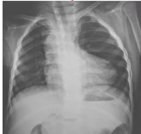
Appeared in: NORCET-7 Mains-2024
Explanation
- The X-ray shows a "boot-shaped heart" or coeur en sabot, which is the classic radiographic sign for Tetralogy of Fallot (TOF).
- This characteristic shape is formed by two key features of TOF: right ventricular hypertrophy, which causes the cardiac apex to be uplifted, and a hypoplastic (underdeveloped) main pulmonary artery, which creates a concave segment on the left heart border.
- The lung fields also show decreased vascular markings (oligemia), consistent with the reduced pulmonary blood flow seen in TOF due to right ventricular outflow tract obstruction.
Why Other Options Were Wrong
- Option A: SAIO is not a recognized medical acronym for a finding on a chest X-ray related to congenital heart disease. It does not describe the visible cardiac anomaly.
- Option B: A diaphragmatic hernia would present with abdominal contents, such as bowel loops (visible as air-filled structures), within the thoracic cavity. This X-ray shows clear lung fields and a well-defined diaphragm without herniated contents.
- Option D: The classic X-ray signs of coarctation of the aorta are rib notching (due to collateral blood flow through intercostal arteries) and the "3 sign" (an indentation of the aorta at the site of coarctation). Neither of these findings is present in the image.
Related Visual

Clinical Relevance
- Nursing practice connection: This is primarily an exam-oriented knowledge point with limited direct bedside application, so retain Radiographic identification of congenital heart defects, specifically Tetralogy of Fallot (TOF) as background academic context rather than a clinical decision trigger.
- Recognizing the boot-shaped heart on a chest X-ray is a critical skill for nurses and clinicians, as it strongly suggests a diagnosis of Tetralogy of Fallot, prompting urgent pediatric cardiology consultation.
- Nurses caring for infants with TOF must be vigilant for signs of hypercyanotic episodes ("Tet spells"), which are life-threatening events characterized by a sudden increase in cyanosis and respiratory distress. Immediate intervention, such as placing the infant in the knee-chest position, is crucial.
- What if? If the X-ray instead showed an "egg-on-a-string" appearance (a large, oval-shaped heart with a narrow superior mediastinum), the most likely diagnosis would be Transposition of the Great Arteries (TGA), another critical cyanotic congenital heart defect requiring different management.
How to Approach the Question
- First, carefully examine the provided image, which is a pediatric chest X-ray. Pay close attention to the size, shape, and position of the heart and great vessels.
- Identify the most prominent abnormal finding. In this case, the heart has a distinct "boot" shape, with an elevated apex and a concave left upper border where the pulmonary artery should be.
- Systematically evaluate the given options. Correlate the visual evidence from the X-ray with the known radiographic signs of each condition.
- Recall or look up the classic X-ray finding for Tetralogy of Fallot (TOF), which is the "boot-shaped heart" or coeur en sabot.
- Eliminate the other options by considering their typical X-ray appearances. A diaphragmatic hernia would show bowel in the chest. Coarctation of the aorta would show rib notching or a "3 sign". SAIO is not a standard diagnosis.
- Conclude that the finding is most consistent with Tetralogy of Fallot based on the classic boot-shaped heart appearance.
Concept Tested & Keywords
- Concept Tested: Radiographic identification of congenital heart defects, specifically Tetralogy of Fallot (TOF).
- Stem keywords: x-ray finding, x-ray film
- Lead-in keywords: Identify
- Clinical cues: Boot-shaped heart on pediatric chest X-ray
Question ID
QRJSiiyj_jTHdTE0vkp2qL
Reference Book
E6 Nelson Textbook of Pediatrics(2024) — Volume 2 p. 588-590
E6 Medicine Davidson Principles Practice 24e p. 470-472
Practise the full NORCET-7 Mains-2024
Attempt every question from this paper in a timed mock, then review the full solution for each one.
More Disorders of Cardiovascular System Questions
- The type of cyanosis seen in PDA is:
- In which disease condition a boot shaped heart is seen in chest X-ray?
- Which of the following is a congenital malformation of the heart?
- Which of the following is NOT a part of the Tetralogy of Fallot (TOF)?
- The congenital heart disease in which radiographic findings depict boot-shaped heart is called?
- Which of the following is the hypertensive Blood Pressure of a 6-year-old child?
- Common position in a child with tetralogy of Fallot?
- Which of the following is a cyanotic heart disease?
- A child is admitted to the pediatric ward with suspected rheumatic arthritis. What should be an important question that a nurse should ask the parent…
- A 1-month-old boy presents with failure to thrive. On examination, he shows features of congestive heart failure. The femoral pulses are feeble as co…
More NORCET-7 Mains-2024 Questions
- What is the correct statement regarding pre-oxygenation in children before intubation?
- A pregnant woman is ready for caesarean section and transferred to labor room. Which is the best position for the woman during caesarean section?
- A woman comes in labor room with 3 cm cervical dilation, 80% effacement and energetic and excited. The women in which phase of labor?
- The ambient temperature of the labor room should maintain?
- The recommended dose of iron and folic acid for adolescent girls under Weekly Iron & Folic Acid Supplementation (WIFS) program?
- A 14-week Rh-negative pregnant woman presents with mild vaginal bleeding and abdominal pain. Ultrasound shows a viable intrauterine pregnancy. Her hu…
- A new newborn care unit is inaugurated in the hospital. The child health nurse known about iKMC. What mean I in iKMC?
- A nurse teaching to a woman regarding breast feeding. Which statement is incorrect about breast feeding?
- Swenson and Duhamel procedure used to correction of?
- Cohort study also known as?