
NORCET-7 Mains-2024
Obstetrics & Gynaecology
Medium
Identify the following image? 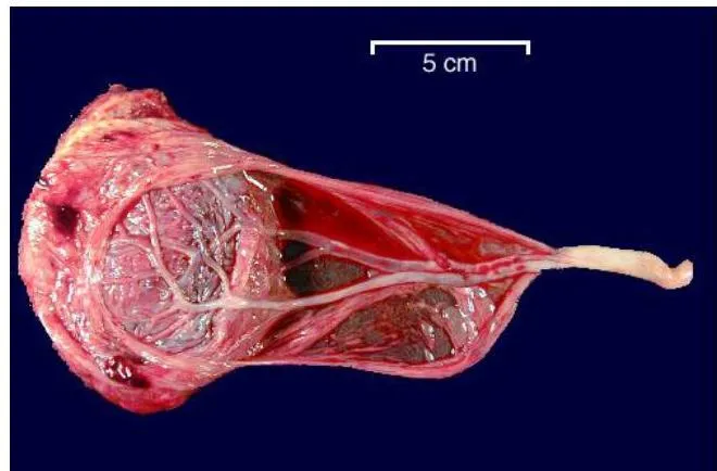
Appeared in: NORCET-7 Mains-2024
Explanation
- The image shows a velamentous insertion of the umbilical cord, a condition where the cord inserts into the fetal membranes instead of the main placental disc.
- As clearly visible in the specimen, the umbilical vessels separate from the cord and travel unprotected through the chorioamniotic membranes to reach the placenta.
- This anatomical variation leaves the fetal vessels vulnerable to compression and rupture, which can lead to fetal hypoperfusion, acidemia, or exsanguination.
- This presentation is distinct from other placental abnormalities like battledore (marginal insertion), succenturiate (accessory lobe), or circumvallate (fibrous ring).
Why Other Options Were Wrong
- Option A: A circumvallate placenta is characterized by a thickened, opaque, white circular ridge on the fetal surface, composed of a double fold of chorion and amnion. This feature is not present in the image.
- Option C: A battledore placenta involves the umbilical cord inserting directly into the margin (edge) of the placental disc. The image shows the cord inserting into the membranes at a distance from the placental edge.
- Option D: A succenturiate lobe is an accessory placental lobe that is separate from the main placental disc, connected by fetal vessels running through the membranes. The image shows a single main placental mass.
Related Visual

- Visual 1: Diagram - A comparative illustration showing a normal central cord insertion alongside velamentous, marginal (battledore), and succenturiate lobe placentas. This helps clarify the anatomical differences.
- Visual 2: Sonogram with Color Doppler - An ultrasound image showing vasa previa, where velamentous vessels are seen crossing the internal cervical os. This highlights the most dangerous complication associated with velamentous insertion.
Clinical Relevance
- Nursing practice connection: This is primarily an exam-oriented knowledge point with limited direct bedside application, so retain Identification of placental abnormalities, specifically variations in umbilical cord insertion as background academic context rather than a clinical decision trigger.
- Nurses must carefully inspect the placenta and cord after delivery to identify abnormalities like velamentous insertion, which may not have been detected prenatally. This finding should be documented and communicated.
- Identification of this condition is crucial as it is associated with higher risks of fetal growth restriction, preterm delivery, low Apgar scores, and stillbirth due to potential vessel compression or rupture.
- What if? If these unprotected vessels were found to be crossing the internal cervical os on a prenatal ultrasound (a condition called vasa previa), management would change significantly. Instead of awaiting spontaneous labor, a planned cesarean delivery, typically between 34 to 36 weeks, would be recommended to prevent catastrophic fetal hemorrhage upon membrane rupture or labor onset.
How to Approach the Question
- First, carefully examine the provided image of the delivered placenta.
- Focus on the key anatomical feature in question: the insertion point of the umbilical cord.
- Observe that the cord does not attach to the main body or edge of the placenta. Instead, it attaches to the surrounding fetal membranes.
- Note the blood vessels branching from the cord and traveling across the membranes before reaching the placental disc. These vessels lack the protection of Wharton's jelly.
- Recall the definitions of the placental abnormalities listed in the options.
- Match your visual finding (cord insertion into membranes with exposed vessels) to the correct definition, which is a velamentous insertion.
Concept Tested & Keywords
- Concept Tested: Identification of placental abnormalities, specifically variations in umbilical cord insertion.
- Stem keywords: placenta, umbilical cord, image identification
- Lead-in keywords: Identify
Question ID
Q3ma1QUp0v3RyBdLpBkexC
Practise the full NORCET-7 Mains-2024
Attempt every question from this paper in a timed mock, then review the full solution for each one.
More Placental Abnormalities Questions
More NORCET-7 Mains-2024 Questions
- What is the correct statement regarding pre-oxygenation in children before intubation?
- A pregnant woman is ready for caesarean section and transferred to labor room. Which is the best position for the woman during caesarean section?
- A woman comes in labor room with 3 cm cervical dilation, 80% effacement and energetic and excited. The women in which phase of labor?
- The ambient temperature of the labor room should maintain?
- The recommended dose of iron and folic acid for adolescent girls under Weekly Iron & Folic Acid Supplementation (WIFS) program?
- A 14-week Rh-negative pregnant woman presents with mild vaginal bleeding and abdominal pain. Ultrasound shows a viable intrauterine pregnancy. Her hu…
- A new newborn care unit is inaugurated in the hospital. The child health nurse known about iKMC. What mean I in iKMC?
- A nurse teaching to a woman regarding breast feeding. Which statement is incorrect about breast feeding?
- Swenson and Duhamel procedure used to correction of?
- Cohort study also known as?