
NORCET 9 Mains - 2025
Medical & Surgical Nursing
Easy
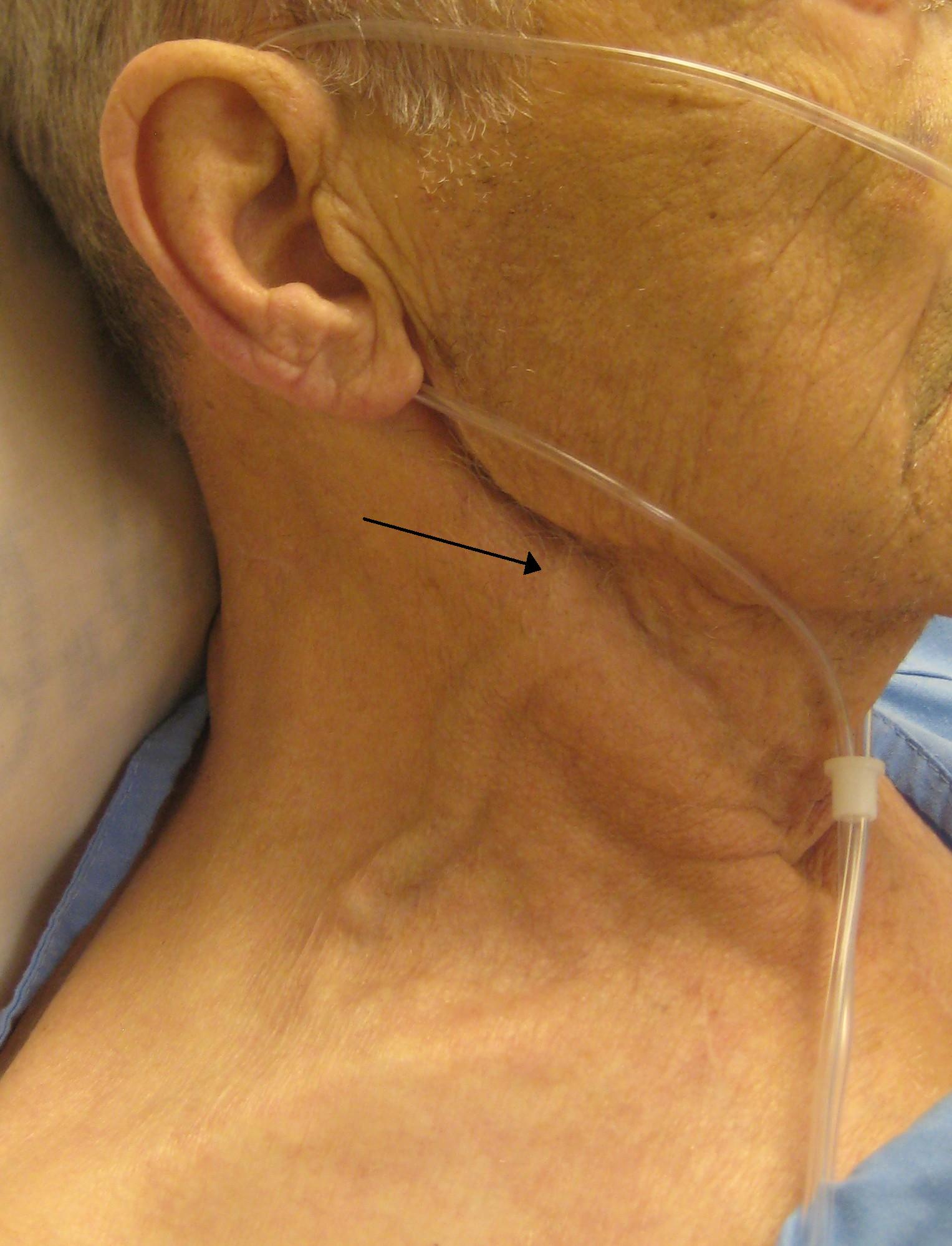
An image shows showing in the following picture. What is the most likely underlying cause?

Appeared in: NORCET 9 Mains - 2025
Explanation
- The image displays Jugular Venous Distension (JVD), a key sign of elevated central venous pressure (CVP).
- Hypervolemia, an excess of fluid within the intravascular space, directly increases CVP by expanding the blood volume.
- This increased pressure and volume cause blood to back up into the jugular veins, leading to the visible distension seen in the neck.
- This finding is crucial for assessing fluid status in conditions like heart failure, kidney injury, or over-infusion of IV fluids.
Why Other Options Were Wrong
- Option A: Dehydration leads to a deficit of total body water and subsequent hypovolemia, causing decreased CVP and flattened neck veins, which is the opposite of JVD.
- Option B: Hypovolemia is a state of decreased circulating blood volume. This condition results in low CVP and would cause the neck veins to be collapsed or flat, not distended.
- Option D: While related, 'fluid overload' is a broader term for excess total body water. Hypervolemia is the more specific term for excess fluid within the blood vessels that directly causes JVD. A patient can have fluid overload with edema (third-spacing) but still be intravascularly depleted.
Related Visual

Clinical Relevance
- Nursing practice connection: This is primarily an exam-oriented knowledge point with limited direct bedside application, so retain Assessment of Fluid Volume Status and Pathophysiology of Jugular Venous Distension as background academic context rather than a clinical decision trigger.
- Assessing for JVD is a fundamental nursing skill for non-invasively estimating a patient's central venous pressure and fluid volume status at the bedside.
- It is a critical indicator for managing conditions like heart failure, guiding diuretic therapy, and preventing complications from excessive IV fluid administration.
- What if? If the patient's neck veins were flat and they had a weak, thready pulse, the most likely cause would shift to Hypovolemia or Dehydration, and the immediate nursing intervention would be fluid resuscitation, not fluid restriction.
How to Approach the Question
- First, identify the key clinical sign shown in the image, which is a distended or bulging neck vein.
- Recognize this sign as Jugular Venous Distension (JVD).
- Recall the underlying pathophysiology: JVD indicates increased pressure and volume in the central venous system.
- Evaluate each option to determine which condition causes increased venous pressure.
- Eliminate options that cause fluid deficit and decreased venous pressure (Dehydration, Hypovolemia).
- Between the remaining similar options (Hypervolemia, Fluid overload), choose the most precise physiological term that directly explains the sign. Hypervolemia specifically refers to the increased intravascular volume causing the distension.
Concept Tested & Keywords
- Concept Tested: Assessment of Fluid Volume Status and Pathophysiology of Jugular Venous Distension
- Stem keywords: jugular vein, underlying cause
- Lead-in keywords: What is the most likely
- Clinical cues: Visual sign of distended neck vein indicates elevated central venous pressure.
Question ID
QBr9K7Q2KAydhywwyuBj_C
Reference Book
E6 Nursing Brunner Adult Health 3SA Vol 1 Part 1 pp. 143-145, 140-142
Practise the full NORCET 9 Mains - 2025
Attempt every question from this paper in a timed mock, then review the full solution for each one.
More Fluid and Electrolytes: Balance and Distribution Questions
- All are crystalloid solution; except:
- ECG of the patient is showing ST depression, Prominent U wave and inverted T wave. Which of the electrolyte imbalance present?
- A patient reports calcium loss. What is the nursing intervention?
- The principle of hemodialysis in which the water is moving under high pressure to an area of lower pressure is:
- Measurement of arterial blood gas shows pH: 7.2, PaCO2 : 70 mm Hg, HCO3: 27 mmol/L and PaO2: 59 mm Hg. Interpret this:
- Fluid volume deficit or hypovolemia describes the loss of
- 100 mL fluid is given to patient and patient's CVP is 3 mm Hg, then what is the next intervention?
- A patient presents to the emergency department with a serum pH of 7.25, pCO2 of 40 mmHg, HCO3 of 22 mEq/L, sodium of 150 mEq/L, and chloride of 100 m…
- A 65-year-old patient is admitted to the hospital with a serum sodium level of 132 mmol/L. Despite being asymptomatic at admission, the patient begin…
- How would you most accurately assess fluid volume status in a patient with 20% total body surface area (TBSA) burns?
More NORCET 9 Mains - 2025 Questions
- A schizophrenia patient believes that the neighbor has installed a circuit inside his body. This is an example of which type of delusion?
- All medical test results are normal, but the patient persistently feels that something is abnormal in the body. This condition is known as:
- Bipolar I disorder is best characterized by:
- Bipolar II disorder is best characterized by:
- A patient taking antipsychotic medication develops extrapyramidal symptoms (EPS). What should be the management?
- A patient develops involuntary, continuous, restless movements after taking antipsychotics. This condition is known as:
- Which drug is the drug of choice in pre-eclampsia?
- PM POSHAN (Pradhan Mantri Poshan Shakti Nirman) scheme was started in which year?
- Which of the following is a killed vaccine?
- Family Planning Programme in India was first started in which year?