
INI-CET EXAM -2026
Pathology & Genetics
Easy
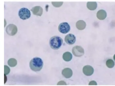
A peripheral blood smear is shown below. Which supravital stain was used?
Appeared in: INI-CET EXAM -2026
Explanation
- New Methylene Blue is a supravital stain, meaning it is applied to living, unfixed cells.
- This stain penetrates immature red blood cells (reticulocytes) and precipitates the residual ribosomal RNA (rRNA) within them.
- The precipitated rRNA forms a characteristic dark blue, net-like structure or 'reticulum', which is visible under a microscope and allows for identification and counting.
- The image clearly shows these reticulocytes with the classic stained reticulum, confirming the use of a supravital stain like New Methylene Blue.
Why Other Options Were Wrong
- Option A: Giemsa stain is a Romanowsky-type stain used for fixed smears to differentiate white blood cells and detect blood parasites like malaria. It does not visualize the reticulum in reticulocytes.
- Option C: Leishman stain is another Romanowsky stain, similar to Giemsa. It is used for differential white blood cell counts and to visualize parasites like Leishmania. It does not stain the reticulocyte network.
- Option D: Diff-Quik is a rapid, modified Romanowsky stain used for quick differential counts in clinical settings. Like other Romanowsky stains, it does not reveal the specific reticulum of reticulocytes.
Related Visual

Clinical Relevance
- Nursing practice connection: Knowing Identification of supravital stains for reticulocyte visualization helps nurses interpret findings accurately and avoid errors in routine assessment, medication administration, and patient teaching.
- The reticulocyte count is a fundamental nursing assessment tool to understand the bone marrow's response to anemia. It helps differentiate between anemias caused by poor production versus those caused by increased destruction or loss of red blood cells.
- A nurse monitoring a patient receiving treatment for iron-deficiency anemia (e.g., iron supplements) would expect to see a rising reticulocyte count as an early sign that the treatment is effective and the bone marrow is starting to produce more RBCs.
- What if? If the smear showed single, eccentric, dark blue dots (not a network) after supravital staining, these would be Heinz bodies. This would shift the diagnosis towards conditions causing oxidative stress on RBCs, such as G6PD deficiency or exposure to certain drugs/toxins.
How to Approach the Question
- First, carefully examine the image provided. Note the presence of red blood cells containing a distinct blue, web-like or granular pattern inside.
- Identify these specific cells as reticulocytes, which are immature red blood cells.
- Read the question stem, which asks to identify the 'supravital stain' used. This term is a major clue, as it refers to a specific category of stains used on living cells.
- Connect the visual finding (reticulocytes) with the key term (supravital stain). The task is to find the specific supravital stain used to visualize reticulocytes.
- Evaluate the options. Recall or deduce that New Methylene Blue is the classic supravital stain for this purpose. Giemsa, Leishman, and Diff-Quik are Romanowsky stains used for different purposes on fixed smears.
Concept Tested & Keywords
- Concept Tested: Identification of supravital stains for reticulocyte visualization.
- Stem keywords: peripheral blood smear, supravital stain
- Lead-in keywords: Which
- Clinical cues: Image showing reticulocytes with a distinct reticulum.
Question ID
Q-dZ4pyt_aQX3VXI-Aatyr
Reference Book
E6 Medicine Harrison 22e Part 1 pp. 478-480, 488-490
E6 Pathology-Vandana Puri & Kavita Gaur Textbook of Pathology and Genetics p. 389-391
Practise the full INI-CET EXAM -2026
Attempt every question from this paper in a timed mock, then review the full solution for each one.
More Clinical Pathology Questions
- Lymphocytes in viral infection?
- In hemophilia A disease bleeding occurs due to the deficiency of?
- Cell with a horseshoe shaped nucleus and abundant cytoplasm in a peripheral blood smear is?
- Cell concentration techniques include all of the following, except?
- Which of the following are not the symptoms of Leukemia?
- The composition of blood transfused to a hemophilia patient is?
- Which type of anemia is characterized by macrocytic RBCs with megaloblastic changes?
- Thalassemia is a congenital?
- Mongoloid faces are seen in children suffering from?
- One of the following disorders in which an abnormal haemoglobin leads to chronic hemolytic anemia with other clinical manifestations due to point mut…
More INI-CET EXAM -2026 Questions
- In the diagram of the abdominal wall musculature, the muscle labelled 'X' and the structure formed by its aponeurosis are best identified as:
- A 45-year-old chronic smoker is being counselled for smoking cessation therapy. Which of the following statements regarding nicotine replacement and…
- Lamotrigine is most effective in which of the following seizure types?& x20;
- In the labelled diagram of laryngeal intrinsic muscles, which labelled muscle is solely responsible for abduction of the vocal cords?
- & x20;Which muscle, as shown in the diagram, attaches to the cartilaginous portion of the Eustachian tube and also blends with the soft palate?
- Q4. Match each of the following organs with its correct embryological tissue of origin: Organ Embryological Origin& x20; A. Thyroid gland 1. Neural c…
- Q5. Match each of the following gastrointestinal organs with its distinctive histological feature: & x20;Organ Histological Feature & x20;1\. Ileum a…
- Q6. A developmental remnant of the embryological hindgut persisting in the retrorectal (presacral) space is most likely to give rise to which of the…
- & x20;The histological section shown in the image, characterized by predominantly serous acini, is best identified as which organ?
- A failure of fusion of the maxillary process with the medial nasal process during embryonic development results in which type of cleft anomaly?